Understanding Brainstem Glioma and Its Types


Brainstem glioma refers to a tumor that develops in the brainstem, a vital part of your central nervous system responsible for controlling essential functions like breathing and heart rate. These tumors are classified based on their location and growth patterns:
Type of Glioma | Growth Characteristics |
|---|---|
Grow slowly, restricted to one area, typically easier to treat, better outcomes. | |
Extremely aggressive, invade neighboring tissues, grow throughout the brainstem. |
This condition is more common in children and young adults.
Brainstem gliomas account for about 20% of brain tumors in children.
In adults, they represent less than 2% of all intracranial tumors.
Understanding these tumors is crucial for early diagnosis and treatment.
Key Takeaways
Brainstem gliomas are growths in the brainstem. They affect important functions like breathing and heartbeat.
There are two main kinds: focal gliomas grow slowly and are easier to treat. Diffuse intrinsic pontine gliomas (DIPG) grow fast and are harder to treat.
Signs of brainstem gliomas include blurry vision, weakness, and trouble eating. See a doctor if you notice these problems.
Finding the tumor early is very important. Tests like MRI help doctors find the tumor and decide on treatment.
Treatments include radiation, chemotherapy, and new experimental methods. Scientists are working to find better ways to help patients.
What Is Brainstem Glioma?
Definition and Overview
Brainstem glioma is a type of tumor that forms in the brainstem, which connects your brain to your spinal cord. This tumor can disrupt essential functions like breathing, heartbeat regulation, and motor control. Brainstem gliomas are rare in adults but more common in children, where they account for a significant percentage of pediatric brain tumors. These tumors can vary in their growth patterns, with some growing slowly and others being highly aggressive.
Why the Brainstem Is a Critical Area
The brainstem plays a vital role in your body’s survival. It controls automatic functions such as breathing, heart rate, and swallowing. It also acts as a communication hub, transmitting signals between your brain and the rest of your body. A tumor in this area can interfere with these critical processes, leading to severe symptoms like difficulty breathing, loss of coordination, or even paralysis. Because of its location, treating a brainstem glioma poses unique challenges.
General Causes and Risk Factors
The exact causes of brainstem gliomas remain unclear. However, genetic mutations, particularly in the histone H3 gene, have been identified as significant contributors. These mutations are especially common in children and suggest that epigenetic changes play a role in the development of diffuse intrinsic pontine glioma (DIPG).
Risk factors for brainstem gliomas differ between children and adults. In children, high-grade tumors like DIPG are more common, with a median survival of about 10 months. Factors such as age under 2 years, cranial nerve palsies, and diffuse intrinsic lesions increase the risk. In adults, low-grade tumors are more frequent, with a median survival ranging from 30 to 85 months. Conditions like neurofibromatosis and symptoms lasting over 12 months are associated with adult cases.
Age Group | Prognosis | Common Tumor Characteristics | Risk Factors |
|---|---|---|---|
Children | Predominantly high-grade tumors, especially DIPG | Age < 2 years, multiple brainstem signs, cranial nerve palsies, diffuse intrinsic lesions | |
Adults | Median survival 30-85 months | Predominantly low-grade tumors | Neurofibromatosis, symptoms lasting > 12 months, exophytic location |
Understanding these factors can help you recognize potential risks and seek early medical attention if needed.
Types of Brainstem Gliomas

Diffuse Intrinsic Pontine Glioma (DIPG)
Definition of DIPG
Diffuse Intrinsic Pontine Glioma (DIPG) is a highly aggressive tumor that forms in the pons, a part of the brainstem. These tumors are classified as grade IV gliomas and are known for their rapid growth and invasive nature. Unlike focal brainstem gliomas, DIPGs spread throughout the brainstem and neighboring structures, making them particularly challenging to treat.
Symptoms of DIPG
DIPG symptoms often develop quickly due to the tumor's aggressive nature. You may notice:
Difficulty with balance and coordination.
Weakness in the arms or legs.
Facial weakness or drooping.
Problems with vision, such as double vision.
Difficulty swallowing or speaking.
Diagnosis of DIPG
Doctors typically diagnose DIPG using imaging techniques like MRI. These scans reveal the tumor's diffuse nature and its location in the pons. Biopsies are rarely performed due to the tumor's critical location and the risks involved.
Treatment Options for DIPG
Treatment focuses on managing symptoms and slowing tumor progression. Radiation therapy is the most common approach, offering temporary relief. Chemotherapy and experimental treatments, such as targeted therapies and clinical trials, are also being explored. Surgery is not an option due to the tumor's invasive nature.
Prognosis for DIPG
DIPG has a poor prognosis. The average survival is about 8-11 months, with only 10% of patients surviving two years and 2% surviving five years.
Survival Rate | |
|---|---|
2-year | ~10% |
5-year | ~2% |
Median Survival | 8-11 months |
DIPG remains one of the most challenging types of brainstem glioma to treat, highlighting the need for continued research and innovation.
Symptoms of Brainstem Gliomas
Common Neurological Symptoms
Brainstem gliomas can cause a wide range of neurological symptoms. These symptoms often result from the tumor pressing on critical areas of the brainstem. You may notice:
Double vision or difficulty focusing.
Weakness in your arms or legs.
An unsteady gait, making walking difficult.
Trouble swallowing food or liquids.
Slurred speech, also known as dysarthria.
Persistent headaches, which are especially common in adults.
Nausea or vomiting, often occurring in the morning.
Drowsiness or unusual fatigue.
Behavioral changes or, in rare cases, seizures in children.
Older children may experience worsening handwriting or speech.
Other signs include cranial nerve deficits, ataxia (loss of coordination in the trunk or limbs), and long tract signs, which indicate damage to motor or sensory pathways. These symptoms can vary in severity depending on the tumor's size and growth rate.
Symptoms Based on Tumor Location
The location of the tumor within the brainstem significantly influences the symptoms you may experience. For example:
Tumors in the pons often cause facial weakness, double vision, and difficulty with balance.
Tumors in the midbrain may lead to problems with eye movement and coordination.
Tumors in the medulla oblongata can affect swallowing, breathing, and heart rate regulation.
Each part of the brainstem controls specific functions, so the symptoms provide clues about the tumor's location. Recognizing these patterns can help doctors pinpoint the affected area during diagnosis.
When to Seek Medical Attention
Certain symptoms should prompt you to seek immediate medical attention. These include:
Persistent double vision or sudden weakness.
Difficulty walking or maintaining balance.
Trouble swallowing or speaking clearly.
Severe headaches that worsen over time.
Nausea or vomiting that doesn’t improve.
Behavioral changes, such as irritability or confusion.
In children, watch for unusual signs like seizures, changes in handwriting, or speech difficulties. Early detection of a brainstem glioma can improve treatment outcomes, so don’t ignore these warning signs.
Diagnosis of Brainstem Gliomas
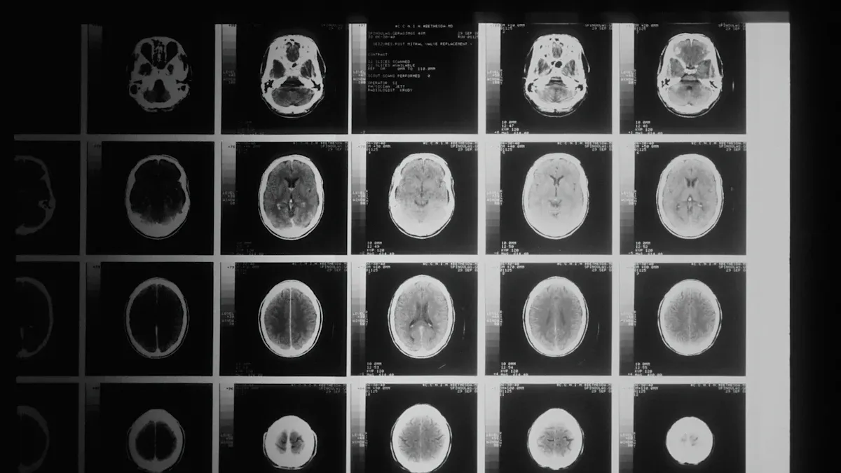
Imaging Techniques (MRI, CT Scans)
Doctors rely on imaging techniques to diagnose brainstem gliomas effectively. Magnetic Resonance Imaging (MRI) is the most accurate tool for identifying these tumors. It provides detailed images of the brainstem, showing diffuse nonenhancing lesions in T1 sequences and hyperintensity in T2-FLAIR sequences. Diffusion Tensor Imaging (DTI) can also help differentiate brainstem gliomas from other conditions like demyelinating diseases. However, Magnetic Resonance Spectroscopy (MRS), which aids in differential diagnosis, faces technical challenges when used in the brainstem.
Imaging Technique | Description |
|---|---|
MRI | Shows diffuse nonenhancing lesions in T1 sequences and hyperintensity in T2-FLAIR sequences. |
MRS | Used for differential diagnosis; limited in brainstem due to technical difficulties. |
Diffusion Tensor Imaging | Helps differentiate diffuse brainstem gliomas from demyelinating diseases. |
When MRI is unavailable, doctors may use CT scans. Although less accurate, CT scans can still provide valuable information about the tumor's location and size.
Biopsy and Histological Analysis
In some cases, doctors may recommend a biopsy to confirm the diagnosis of a brainstem glioma. This procedure involves removing a small sample of the tumor tissue for histological analysis. The analysis helps determine the tumor's grade and type, which guides treatment decisions. However, biopsies in the brainstem carry significant risks due to the area's critical functions. For this reason, doctors often rely on imaging alone for diagnosis, especially in children.
Challenges in Diagnosing Brainstem Gliomas
Diagnosing brainstem gliomas presents several challenges. MRI remains the diagnostic test of choice, but tissue confirmation through biopsy is sometimes unfeasible due to the tumor's location. CT scans, while useful in certain situations, are less accurate than MRI.
Studies show that preoperative radiological diagnoses can be incorrect in 10%–25% of cases for patients over 20 years old with contrast-enhancing lesions. In some instances, benign non-neoplastic pathologies were mistaken for malignant brainstem masses.
These challenges highlight the importance of combining advanced imaging techniques with clinical expertise to ensure an accurate diagnosis.
Treatment Options and Prognosis
Treatment Approaches
Radiation Therapy
Radiation therapy remains the cornerstone of treatment for brainstem gliomas. Focal radiotherapy, with doses ranging from 54 to 60 Gy, can stabilize or improve symptoms in many patients. It is particularly effective for those with progressive neurological symptoms. However, its benefits are often temporary, and the tumor may eventually progress. Patients with exophytic tumors tend to have better outcomes compared to those with diffuse intrinsic lesions.
Evidence Type | Description |
|---|---|
Treatment Role | Radiotherapy is the standard treatment for adult diffuse intrinsic gliomas. |
Clinical Improvement | 61% of patients show clinical improvement, though radiological response is lower (19%). |
Chemotherapy
Chemotherapy plays a secondary role in treating brainstem gliomas. Drugs like temozolomide and carboplatin/vincristine are commonly used. However, their effectiveness is limited, with only modest clinical improvements observed in some cases. Antiangiogenesis agents, such as bevacizumab, are being studied, but their role remains unclear.
Study Focus | Findings |
|---|---|
Chemotherapy Response | Modest radiological response (10%). |
Clinical Improvement | Noted in 18% of cases. |
Overall Efficacy | Adjuvant chemotherapy not recommended. |
Surgery (When Applicable)
Surgical resection is rarely an option due to the brainstem's critical functions. However, it may be considered for specific cases, such as tumors at the cervicomedullary junction or cystic tumors. These cases allow for safer removal with minimal risk to vital structures.
Experimental Treatments and Clinical Trials
Experimental treatments offer hope for patients with brainstem gliomas. Clinical trials explore innovative therapies, including immune system activation and new drug combinations. For example, IDH inhibitors have shown promise in slowing tumor progression in certain gliomas. Enrolling in clinical trials provides access to cutting-edge treatments that may improve outcomes.
Prognosis and Factors That Influence Outcomes
The prognosis for brainstem gliomas depends on several factors. Favorable indicators include low-grade tumor histology, exophytic location, and symptoms lasting over 12 months before diagnosis. Poor prognostic factors include diffuse intrinsic lesions, cranial nerve palsies, and high-grade histology.
Favorable Prognostic Factors | Poor Prognostic Indicators |
|---|---|
Neurofibromatosis | Age younger than 2 years |
Symptoms lasting ≥12 months | Multiple brainstem signs |
Exophytic location | Cranial nerve palsies |
Coping and Support for Patients and Families
Coping with a brainstem glioma diagnosis can be overwhelming. Support resources, such as caregiver programs, psycho-oncology services, and survivor support groups, can help you navigate this journey. Nutrition counseling and exercise programs also play a role in maintaining overall well-being. Families may benefit from pediatric support services and palliative care to address emotional and physical needs.
Coping Strategy | Link |
|---|---|
Caregiver Program | |
Support Groups | |
Palliative Care |
Brainstem glioma remains a challenging condition, but understanding its types and symptoms can help you recognize the importance of early diagnosis. Identifying the tumor’s location and characteristics allows doctors to tailor treatment strategies, improving outcomes. Exploring options like radiation therapy, chemotherapy, and experimental treatments offers hope for managing this condition.
Ongoing research continues to advance treatment possibilities. For example:
Advancement | Description |
|---|---|
Tumor Treating Fields (TTF) Therapy | Uses electrical fields to disrupt cancer cell multiplication with minimal side effects. |
IDH Inhibitors | Targeted therapies that may slow the progression of low-grade gliomas. |
Immune Therapy Combinations | Clinical trials are exploring ways to activate the immune response against gliomas. |
Senolytic Drugs | Investigating drugs that kill senescent cells to prevent tumor recurrence. |
These advancements highlight promising directions:
Immune therapies face challenges but show potential in clinical trials.
Senolytic drugs offer a novel approach to managing residual tumor cells.
With continued research, you can remain hopeful for improved treatments and outcomes.
FAQ
What is the difference between focal brainstem glioma and DIPG?
Focal brainstem gliomas grow slowly and stay localized, making them easier to treat. DIPG, on the other hand, spreads aggressively throughout the brainstem, making treatment more challenging.
Can brainstem gliomas be cured?
Most brainstem gliomas cannot be completely cured. Treatments like radiation therapy and experimental options aim to manage symptoms and slow tumor growth.
Are brainstem gliomas hereditary?
Brainstem gliomas are not typically hereditary. However, genetic mutations, such as those in the histone H3 gene, play a significant role in their development.
How long does it take to diagnose a brainstem glioma?
Diagnosis depends on symptom severity and access to imaging tools like MRI. In most cases, doctors can confirm the diagnosis within a few weeks after symptoms appear.
What should you do if you suspect a brainstem glioma?
Seek medical attention immediately if you notice symptoms like persistent headaches, double vision, or difficulty walking. Early diagnosis improves treatment options and outcomes.
See Also
An In-Depth Overview of Various Cancer Types
Exploring Cancer Types Associated With AIDS Infection
Understanding Angioimmunoblastic T-Cell Lymphoma and Its Signs

© 2026 Banish Cancer. Banish Cancer is a registered non-profit organization providing evidence-based research, educational courses, and Fear Response AI support services. Reg. No: 305706884 | Address: Taikos pr. 43-603, Kaunas, Lithuania.