Do Mammograms Cause Cancer? The Truth Revealed

You might wonder, "Do Mammograms Cause Cancer? Debunking the Radiation Myth," as concerns about radiation exposure arise. The truth is, the radiation dose from a mammogram is extremely low. For perspective:
A mammogram delivers about 0.4 mSv of radiation.
A chest CT scan exposes you to 7 mSv.
PET/CT scans can go as high as 25 mSv.
This makes mammograms one of the safest imaging methods available. Health experts agree that the benefits of early detection far outweigh the risks. Studies show that regular screenings significantly reduce breast cancer deaths. For example, a 10-year study found that cancers caught early through mammograms had better survival rates and lower relapse risks.
Early detection saves lives. Mammograms provide a powerful tool to catch cancer early, giving you the best chance for effective treatment.
Key Takeaways
Mammograms use low radiation, like 26 days of natural exposure. They are safe for regular use.
Finding cancer early with mammograms lowers death rates by 15% to 20%. This helps save lives.
Regular checks mean less harsh treatments and better results for patients.
Experts say women should start mammograms at age 40. Women in their 40s should get them yearly, and women 50 to 74 every two years.
Talk to your doctor about any worries. Make a screening plan that fits your health.
Do Mammograms Cause Cancer? Debunking the Radiation Myth
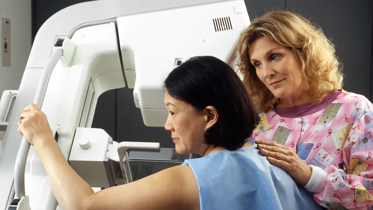
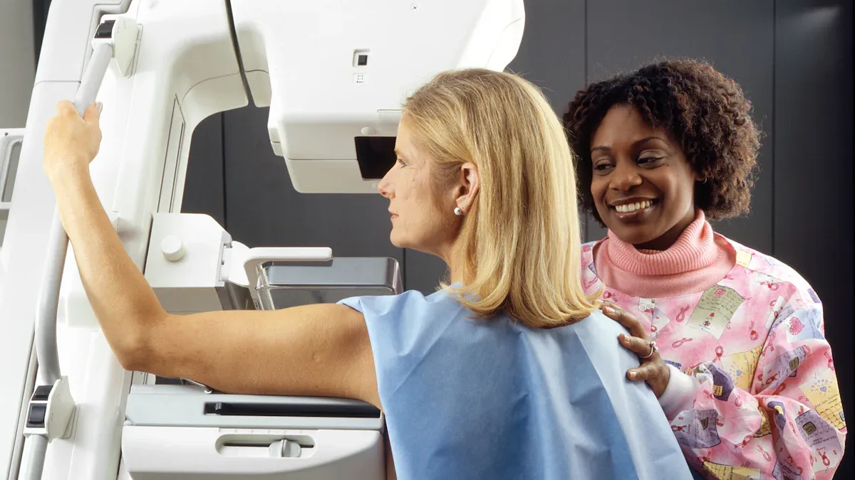
How Mammograms Work
Mammograms use low-dose X-rays to create detailed images of your breast tissue. During the procedure, a technician positions your breast on a flat surface and compresses it gently with a paddle. This compression spreads the tissue, allowing the X-ray to capture clear images. These images help doctors detect abnormalities, such as lumps or calcifications, that might indicate cancer.
The process is quick and non-invasive. It typically takes about 20 minutes, and the X-rays used are specifically calibrated to minimize radiation exposure while providing accurate results.
Radiation Levels in Mammograms
Comparison to everyday radiation exposure
The radiation from a mammogram is minimal. For perspective, it’s comparable to the amount of radiation you’d receive during a transatlantic flight. To illustrate this further:
Measurement Type | Radiation Level (mSv) | Equivalent Background Exposure Time |
|---|---|---|
Mammogram | 7 weeks |
This level is far below what you encounter in other medical imaging tests, such as CT scans.
Why the levels are considered safe by medical standards
The radiation dose from a mammogram equals about 26 days of natural background radiation. Even with annual screenings, the exposure remains well within safe limits set by health organizations. Experts agree that the benefits of early detection far outweigh this negligible risk.
Safety Standards for Mammograms
Guidelines set by health organizations
Health organizations like the FDA and the American Cancer Society ensure mammograms meet strict safety standards. These guidelines regulate equipment and radiation levels to protect you during screenings.
Regular monitoring to ensure safety
Facilities offering mammograms undergo regular inspections to maintain compliance with safety protocols. This ensures that the equipment functions correctly and radiation exposure stays within approved limits.
The question "Do Mammograms Cause Cancer? Debunking the Radiation Myth" often arises, but the evidence overwhelmingly supports their safety.
Scientific Evidence on Mammograms and Cancer Risk
Research Studies
Studies showing no link between mammograms and cancer
You might wonder if mammograms could increase your cancer risk. Research consistently shows that mammograms are safe and effective for detecting breast cancer. The National Cancer Institute highlights that while radiation exposure is a potential concern, the benefits of early detection far outweigh the risks.
Mammograms use low-dose radiation, which typically does not cause harm.
A 2016 study confirmed that although repeated mammography might slightly increase breast cancer risk, the overall benefits of screening are significant.
Regulatory agencies like the FDA ensure that radiation levels during mammograms remain within safe limits.
Long-term data supporting mammogram safety
Long-term studies provide further reassurance about mammogram safety. The table below compares the radiation exposure from different types of mammograms to natural background radiation:
Type of Mammogram | Equivalent Natural Background Radiation | Conclusion |
|---|---|---|
Digital Mammography | 26 days | Low radiation exposure does not significantly increase cancer risk. |
Digital Breast Tomosynthesis (3D) | 33 days | Benefits of early detection outweigh potential risks. |
Recent reviews also confirm that the advantages of early detection surpass the minimal risks associated with radiation exposure.
Expert Opinions
Statements from oncologists and radiologists
Experts agree that mammograms save lives. Oncologists and radiologists emphasize that the benefits of early detection far exceed the negligible risks of radiation. For example:
The sensitivity of mammography ranges from 70% to 90%, depending on factors like age and breast density.
On average, mammograms detect 80% of breast cancers, making them a reliable screening tool.
Recommendations from health organizations
Health organizations like the American Cancer Society (ACS) strongly recommend regular mammograms. Their systematic review found that screening mammography reduces breast cancer mortality.
Randomized controlled trials showed a relative risk (RR) of 0.80–0.82.
Cohort studies reported an RR of 0.75, with a 95% confidence interval (CI) of 0.69–0.81.
Modeling studies estimated a median RR of 0.85, ranging from 0.77 to 0.93.
These findings highlight the life-saving potential of mammograms, reinforcing their importance in breast cancer prevention.
The Benefits of Mammograms
Early Detection Saves Lives
How early detection improves survival rates
Mammograms play a critical role in detecting breast cancer early, often before symptoms appear. Early detection significantly improves survival rates. Studies show that regular screenings reduce breast cancer mortality by 15% to 20%. This means more lives are saved when cancer is caught at an earlier stage.
Statistic Description | Value |
|---|---|
Relative improvement in breast cancer mortality | 15% to 20% |
Absolute improvement at the individual level | Much less than relative improvement |
Lives extended due to early detection | Expressed as a number of lives extended |
Screen-detected cancers also have better outcomes compared to cancers found later. For example, women with screen-detected cancers have a lower relative risk of death compared to those with interval or symptomatic cancers.
Study Description | Relative Risk (RR) for Death | Confidence Interval (CI) |
|---|---|---|
Interval and incident cancers vs. screen-detected cancers | 1.53 | 1.17–2.00 |
Symptomatic women vs. screen-detected cancers | 0.79 | 0.63–0.99 |
Examples of cancers caught early through mammograms
Mammograms can detect changes in breast tissue, such as small lumps or calcifications, long before they become noticeable. Women who undergo regular screenings often discover cancers at stage 0 or stage I, where treatment is most effective. These early-stage detections lead to higher survival rates and better quality of life.
Reducing the Need for Aggressive Treatments
Early-stage cancers often require less invasive treatments
When mammograms detect cancer early, treatments tend to be less aggressive. Early-stage cancers often require smaller surgeries, such as lumpectomies, instead of mastectomies. Chemotherapy may also be avoided in some cases.
Comparison Type | Early-Stage Cancers | Advanced Cancers | Hazard Ratio (HR) | Relative Risk (RR) |
|---|---|---|---|---|
Screen-detected vs. non-screened | Better prognosis | Worse prognosis | 1.90 (95% CI, 1.15–3.11) | 1.53 (95% CI, 1.17–2.00) |
Women with advanced cancers often face more invasive treatments, such as extensive surgeries or prolonged chemotherapy. Early detection helps you avoid these challenges and improves your overall prognosis.
Peace of Mind for Patients
Regular screenings provide reassurance
Mammograms not only save lives but also offer peace of mind. Knowing that you are taking proactive steps to monitor your health can reduce anxiety. Regular screenings help you stay informed about your breast health, even if no abnormalities are found. This reassurance empowers you to focus on other aspects of your well-being.
Early detection through mammograms gives you control over your health and increases your chances of a positive outcome.
Addressing Common Concerns
Myths About Mammograms
"Radiation from mammograms causes cancer."
You might have heard that the radiation from mammograms can cause cancer. This is a common myth. Mammograms use low-dose X-rays, exposing you to minimal radiation. For comparison, the radiation from a single mammogram equals about 26 days of natural background radiation. Regulatory agencies like the FDA ensure that mammogram radiation levels remain within safe limits.
To put this into perspective:
Radiation Exposure Level | Estimated Cancer Risk |
|---|---|
1 in 2,000 | |
Below 10 mSv | Insufficient data for conclusions |
The FDA estimates that 10 mSv of radiation increases lifetime cancer risk by only 1 in 2,000. Since mammograms involve much lower doses, the risk is negligible.
"Frequent mammograms increase cancer risk."
Another misconception is that frequent mammograms increase cancer risk. However, the cumulative dose from regular screenings over 35 years is about 84 mGy, which is still far below harmful levels. Radiation does not accumulate in your body like water in a glass. Instead, your body processes and eliminates it.
Concerns About Repeated Exposure
Why annual or biennial screenings are safe
Annual or biennial mammograms are safe because the radiation dose remains minimal. For example, biennial screenings between ages 50 and 74 are predicted to cause only 7.7 breast cancers and 1.6 breast cancer deaths per 100,000 women. At the same time, these screenings prevent 1,121 breast cancer deaths.
Screening Type | Average Absorbed Dose (mGy) | Total Glandular Dose (mGy) |
|---|---|---|
Biennial (50-74) | 1.3 per view | 18.2 |
Annual (40-49) | Varies | Justified if mortality reduction is ≥ 20% |
The benefits of early detection far outweigh the negligible risks of repeated exposure.
Alternatives to Mammograms
Other screening methods and their limitations
While mammograms are the gold standard, other screening methods exist. These include breast ultrasounds, MRIs, and thermography. However, each has limitations:
Ultrasounds: Useful for dense breast tissue but less effective at detecting small calcifications.
MRIs: Highly sensitive but expensive and prone to false positives.
Thermography: Lacks sufficient evidence to detect cancer reliably.
Study Type | Sample Size | Findings | AUC (Film) | AUC (Digital) |
|---|---|---|---|---|
Dutch Study | 6 million | Higher recall and detection for digital | 0.92 | 0.91 |
Meta-analysis | 82,573 | No significant difference in detection | 0.92 | 0.91 |
Mammograms remain the most effective tool for early detection, offering a balance of accuracy, accessibility, and safety.
Mammograms are safe and effective tools for early cancer detection. The National Cancer Institute highlights several key benefits:
Early detection through mammograms reduces breast cancer mortality rates.
The radiation exposure is minimal, even lower than standard X-rays.
Regular screenings lead to less aggressive treatments and higher cure rates.
Annual screenings starting at age 40 prevent more deaths than they cause. For women aged 50 to 74, screenings save over 1,100 lives per 100,000 women while posing negligible risks. Consult your healthcare provider to create a screening plan that works best for you.
FAQ
What age should you start getting mammograms?
Most health organizations recommend starting mammograms at age 40. However, your doctor might suggest earlier screenings if you have a family history of breast cancer or other risk factors. Always consult your healthcare provider for personalized advice.
How often should you get a mammogram?
For women aged 50 to 74, biennial screenings are typically recommended. Women in their 40s may benefit from annual screenings. Your doctor can help determine the best schedule based on your health and risk factors.
Are mammograms painful?
You might feel slight discomfort during the procedure due to breast compression. This step ensures clear images. The sensation is temporary and usually mild. If you experience significant pain, inform the technician so adjustments can be made.
Can mammograms detect all types of breast cancer?
Mammograms are highly effective but not perfect. They may miss some cancers, especially in dense breast tissue. Additional tests like ultrasounds or MRIs can complement mammograms for more accurate detection in certain cases.
Is there a risk of false positives with mammograms?
Yes, false positives can occur, especially in younger women or those with dense breasts. This means the test might suggest cancer when none exists. Follow-up tests help confirm results and ensure accurate diagnosis.
Tip: Discuss any concerns about mammograms with your doctor to make informed decisions about your health.
See Also
Exploring The Signs And Origins Of Breast Cancer
Identifying The Signs Of Breast Cancer In Men
Examining The Symptoms And Triggers Of Bladder Cancer

© 2026 Banish Cancer. Banish Cancer is a registered non-profit organization providing evidence-based research, educational courses, and Fear Response AI support services. Reg. No: 305706884 | Address: Taikos pr. 43-603, Kaunas, Lithuania.