What Is Extrahepatic Bile Duct Cancer Explained Simply

Extrahepatic bile duct cancer is a rare disease that forms in the bile ducts outside your liver. These ducts play a vital role in moving bile, a fluid that helps digest fats, from your liver to your small intestine. When cancer develops here, it can block the flow of bile, leading to serious health problems.
Early detection significantly impacts survival rates. For example, if the cancer is localized or regional at diagnosis, the five-year survival rate is 18%. However, this drops to just 2% when the cancer spreads to distant parts of the body. Understanding this condition can help you recognize symptoms early and seek timely treatment.
Key Takeaways
Extrahepatic bile duct cancer is uncommon but dangerous. Spotting symptoms like yellow skin or belly pain helps early diagnosis.
Knowing the difference between perihilar and distal bile duct cancers is important. Each type has its own traits and treatments.
Finding the cancer early can save lives. If caught early, 18 out of 100 people survive five years. If it spreads, only 2 out of 100 survive.
Watch for risks like liver problems or being overweight. Lowering these risks can reduce your chances of getting this cancer.
If you see symptoms, visit a doctor quickly. Acting fast can lead to better treatment and recovery.
Understanding Extrahepatic Bile Duct Cancer
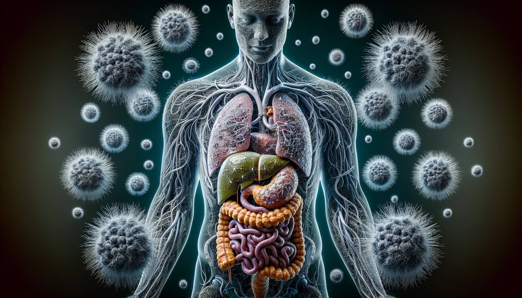
What Are Bile Ducts and Their Function?
Bile ducts are small tubes in your body that carry bile, a digestive fluid produced by your liver. Bile helps break down fats in the food you eat, making it easier for your body to absorb nutrients. These ducts form a network, starting inside the liver and extending outside it. The extrahepatic bile ducts, located outside the liver, connect to your small intestine, where bile mixes with food to aid digestion. Without properly functioning bile ducts, your body struggles to process fats, leading to digestive issues and other health problems.
The Difference Between Intrahepatic and Extrahepatic Bile Ducts
You might wonder how intrahepatic and extrahepatic bile ducts differ. Intrahepatic bile ducts are located inside the liver, while extrahepatic bile ducts are outside it. The table below highlights their structural differences:
Feature | Intrahepatic Bile Ducts | Extrahepatic Bile Ducts |
|---|---|---|
Composition | Interconnected ductules lined by hepatocytes and cholangiocytes | Composed of larger ducts including hepatic and common bile ducts |
Epithelial Morphology | Cuboidal epithelium in interlobular ducts, cylindrical in septal ducts | Varies, typically more robust epithelium |
Associated Structures | Accompanied by branches of hepatic artery and portal vein | Gallbladder attached via loose connective tissue |
Understanding these differences is crucial because intrahepatic bile duct cancer starts inside the liver and may resemble liver cancer. In contrast, extrahepatic bile duct cancer occurs outside the liver and includes perihilar and distal types. These distinctions influence how doctors diagnose and treat each condition.
Why Is Extrahepatic Bile Duct Cancer Significant?
Extrahepatic bile duct cancer is rare, which makes it harder to recognize and diagnose. Certain conditions, like colitis or specific liver diseases, increase your risk of developing this cancer. Symptoms such as jaundice and abdominal pain can severely affect your quality of life. Unfortunately, these signs often appear in advanced stages, making early detection challenging. This highlights the importance of raising awareness about extrahepatic bile duct cancer to improve timely diagnosis and treatment options.
Types of Extrahepatic Bile Duct Cancer
Perihilar Bile Duct Cancer (Klatskin Tumor)
Location and Characteristics
Perihilar bile duct cancer, also known as Klatskin tumor, develops at the junction where the right and left bile ducts meet outside the liver. This type accounts for the majority of extrahepatic bile duct cancer cases. It often grows slowly, making early detection difficult. You might notice symptoms like jaundice or itching as the tumor blocks bile flow.
Doctors classify this cancer into stages based on how far it has spread. The table below outlines these stages:
Stage | Description |
|---|---|
Stage 0 | Abnormal cells are found in the innermost layer of tissue lining the perihilar bile duct. |
Stage I | Cancer has formed in the innermost layer and has spread into the muscle or fibrous tissue layer. |
Stage II | Cancer has spread through the wall to nearby fatty tissue or liver tissue. |
Stage IIIA | Cancer has spread to branches on one side of the hepatic artery or portal vein. |
Stage IIIB | Cancer has spread to the main part of the portal vein or its branches on both sides. |
Stage IIIC | Cancer has spread to one to three nearby lymph nodes. |
Stage IVA | Cancer has spread to four or more nearby lymph nodes. |
Stage IVB | Cancer has spread to other parts of the body, such as the liver, lung, or distant lymph nodes. |
How It Differs from Other Types
Perihilar bile duct cancer stands out because of its location and slow progression. Unlike distal bile duct cancer, which occurs closer to the small intestine, perihilar tumors grow near the liver. This proximity to vital structures makes surgical removal more challenging. You may also experience symptoms earlier with perihilar cancer due to its tendency to block bile flow at a critical junction.
Distal Bile Duct Cancer
Location and Characteristics
Distal bile duct cancer forms in the section of the bile duct closer to the small intestine. This type is less common than perihilar cancer but still significant. You might hear doctors refer to its earliest stage as carcinoma in situ (CIS). The cancer progresses through stages, from stage I (localized) to stage IV (advanced). The TNM staging system evaluates tumor size, lymph node involvement, and metastasis.
Key features of distal bile duct cancer include:
The earliest stage is stage 0, known as carcinoma in situ (CIS).
Stages range from stage I (1) to stage IV (4), with lower numbers indicating less spread.
The TNM staging system assesses tumor size, lymph node involvement, and metastasis.
Each stage may have subcategories (A, B) for more detailed classification.
How It Differs from Other Types
Distal bile duct cancer differs from perihilar cancer in its location and symptoms. Because it occurs closer to the small intestine, you might experience digestive issues or abdominal pain earlier. Treatment options also vary. Surgery is often more feasible for distal tumors due to their location, making early diagnosis crucial for better outcomes.
Symptoms and Risk Factors of Extrahepatic Bile Duct Cancer

Common Symptoms to Watch For
You should pay attention to specific symptoms that might indicate extrahepatic bile duct cancer. These symptoms often arise when the bile duct becomes blocked or inflamed:
Jaundice: Yellowing of the skin and eyes, often the first noticeable sign.
Itching: Caused by excess bilirubin in the skin.
Light-colored or greasy stools: A result of bile not reaching the intestines.
Dark urine: Due to high bilirubin levels in the blood.
Abdominal pain: Often felt in the upper right side of the abdomen.
Loss of appetite and weight loss: Common in many patients.
Fever: Some individuals may experience this symptom.
These symptoms differ from other gastrointestinal conditions, which might present with diarrhea or general abdominal discomfort. If you notice these signs, consult a healthcare provider promptly.
Risk Factors That Increase Susceptibility
Certain factors can increase your risk of developing extrahepatic bile duct cancer. These include:
Liver fluke infections and abnormal bile duct anatomy.
Cirrhosis, hepatitis B or C, and inflammatory bowel disease.
Genetic disorders and older age.
Ethnicity and geography, particularly in regions where liver fluke infections are common.
Obesity, diabetes, and alcohol consumption.
Exposure to harmful substances like Thorotrast, asbestos, and radon.
Other factors such as smoking, chronic pancreatitis, and HIV infection.
Understanding these risk factors can help you assess your susceptibility and take preventive measures.
The Importance of Early Detection and Diagnosis
Early detection plays a critical role in managing extrahepatic bile duct cancer. Imaging techniques like ultrasound, CT scans, and MRCP can help identify abnormalities. However, endoscopic retrograde cholangiopancreatography (ERCP) offers a more precise method. ERCP allows doctors to examine the bile ducts directly and collect tissue samples for analysis. This approach improves the chances of detecting cancer in its early stages.
By recognizing symptoms early and understanding your risk factors, you can seek timely medical advice. Early diagnosis increases the likelihood of successful treatment and better outcomes.
Extrahepatic bile duct cancer is a rare but serious condition that demands your attention. Recognizing its types, symptoms, and risk factors can help you act quickly. Early detection improves survival rates, especially when you understand the differences between perihilar and distal bile duct cancers. By staying informed, you can take proactive steps toward timely diagnosis and treatment. Raising awareness about this condition not only benefits you but also helps others who may face similar challenges. Knowledge truly makes a difference in managing this disease.
FAQ
What is the survival rate for extrahepatic bile duct cancer?
The survival rate depends on the stage at diagnosis. If detected early, the five-year survival rate is around 18%. However, it drops to 2% when the cancer spreads to distant areas. Early detection improves your chances significantly.
Can extrahepatic bile duct cancer be prevented?
You cannot completely prevent it, but reducing risk factors helps. Avoid smoking, maintain a healthy weight, and manage conditions like hepatitis or cirrhosis. Regular check-ups can also aid in early detection.
How is extrahepatic bile duct cancer diagnosed?
Doctors use imaging tests like CT scans, MRCP, or ERCP. ERCP allows direct examination of the bile ducts and tissue sampling. These methods help identify abnormalities and confirm the diagnosis.
Is surgery always an option for treatment?
Surgery depends on the tumor's location and stage. Early-stage cancers are more likely to be operable. Advanced stages may require other treatments like chemotherapy or radiation.
What should you do if you notice symptoms?
If you experience jaundice, itching, or abdominal pain, consult a healthcare provider immediately. Early medical attention increases the likelihood of successful treatment and better outcomes.
Tip: Keep track of any unusual symptoms and share them with your doctor. Early action can save lives.
See Also
Essential Insights About Carcinoid Tumors You Must Have
Key Features and Insights on Cholangiocarcinoma Explained
Recognizing Symptoms and Treatment Options for Duodenal Cancer

© 2026 Banish Cancer. Banish Cancer is a registered non-profit organization providing evidence-based research, educational courses, and Fear Response AI support services. Reg. No: 305706884 | Address: Taikos pr. 43-603, Kaunas, Lithuania.