What is Fibrocartilaginous Mesenchymoma of Bone

Fibrocartilaginous mesenchymoma of bone is a rare type of tumor that develops in the bones. It accounts for less than 1% of all bone tumors worldwide, highlighting its uncommon occurrence. This tumor primarily affects children and adolescents, often appearing in the long bones. Its locally aggressive nature stems from several factors, including rapid growth, the ability to infiltrate nearby tissues, and destruction of the surrounding cortical bone. Histologically, it features spindle cells, dense fibrous stroma, and cartilage resembling the epiphyseal plate. Without timely intervention, it can cause significant damage to the affected area.
Key Takeaways
Fibrocartilaginous mesenchymoma is a very rare bone tumor. It mostly affects kids and young adults. Finding it early helps with better treatment.
Usual symptoms are pain, swelling, and stiffness in the area. Spotting these signs early can help with quick treatment.
Surgery is the main way to treat this tumor. Removing the whole tumor lowers the chance of it coming back. It also helps patients feel better in the long run.
Regular check-ups and imaging tests are very important. These help doctors watch for the tumor returning. Being careful can improve how patients live.
New research on special treatments looks hopeful for the future. This shows why improving medical care is so important.
Understanding Fibrocartilaginous Mesenchymoma of Bone
What is Fibrocartilaginous Mesenchymoma of Bone?
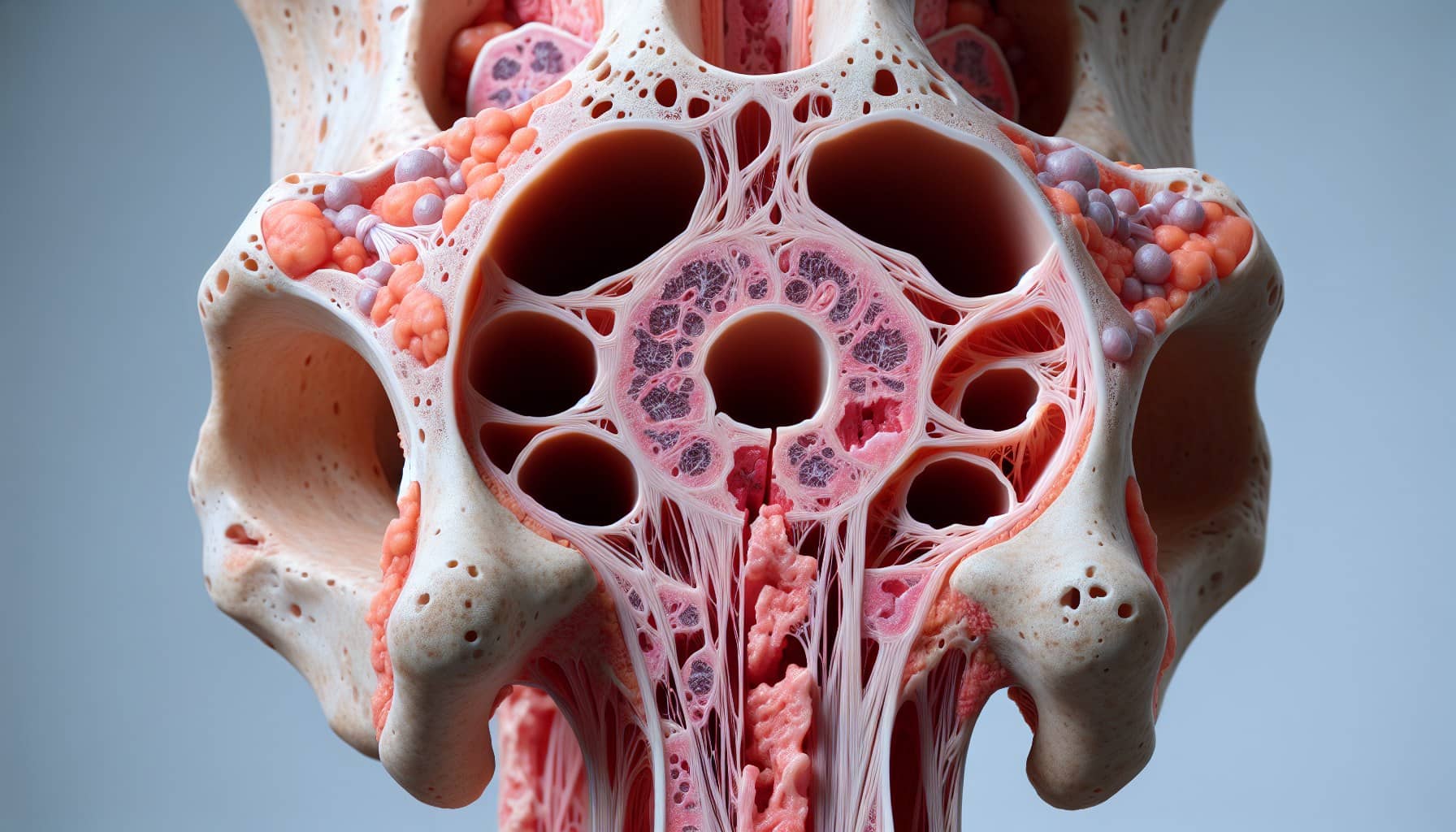
Fibrocartilaginous mesenchymoma of bone is a rare tumor that primarily affects the skeletal system. It is histologically unique, featuring a combination of spindle cells, hyaline cartilage nodules, and bone trabeculae. The spindle cells often display mild cytological atypia, with elongated shapes and slightly hyperchromatic nuclei. These cells are arranged in bundles or intersecting fascicles. The cartilaginous nodules resemble growth plate cartilage, with chondrocytes aligned in parallel columns. Additionally, areas of enchondral ossification and clusters of osteoclast-type giant cells may be present. This tumor can infiltrate the surrounding bone and soft tissues, making early diagnosis crucial.
Key Characteristics of the Tumor
Fibrocartilaginous mesenchymoma of bone stands out due to its distinct histological features. Unlike other bone tumors, it combines spindle cells with mild atypia, cartilage nodules, and bone trabeculae. The cartilaginous component mimics the epiphyseal growth plate, while the spindle cells show moderate cellularity and mild hyperchromasia. This tumor can grow rapidly and reach significant sizes, despite being classified as benign. Differential diagnosis is essential, as it can be mistaken for conditions like fibrous dysplasia or low-grade fibrosarcoma. However, the absence of high-grade sarcomatous components and the presence of cartilage nodules help distinguish it from other tumors.
Who is Most Commonly Affected?
Fibrocartilaginous mesenchymoma of bone predominantly affects children and young adults. Most cases occur between the ages of 10 and 30, with a median age of 13 years. The youngest reported patient was just three months old, while the oldest was 27 years. Males have a slightly higher incidence compared to females. This tumor is extremely rare, with fewer than 40 documented cases worldwide. Its rarity and unique presentation make it a challenging condition for clinicians to diagnose and manage effectively.
Clinical Features and Symptoms
Common Symptoms of Fibrocartilaginous Mesenchymoma
Fibrocartilaginous mesenchymoma of bone often presents with a range of symptoms that vary depending on the tumor's size and location. Commonly reported symptoms include:
Swelling in the surrounding area
Stiffness in the nearby joint
Restricted range of motion
In some cases, the tumor may remain asymptomatic for a period
Pain and swelling are the most frequent complaints, particularly in weight-bearing bones. These symptoms can interfere with daily activities, especially in younger patients who are more active. Early recognition of these signs is essential for timely diagnosis and treatment.
How the Tumor Presents Clinically
Clinically, fibrocartilaginous mesenchymoma may appear as either a painful lesion or a localized swelling. Patients often report persistent pain in the affected bone or joint, which may worsen over time. Swelling and stiffness in the area can further limit mobility. In some cases, the tumor may restrict the range of motion in nearby joints, making movement difficult. While some individuals experience significant discomfort, others may remain asymptomatic until the tumor grows larger. This variability in presentation highlights the importance of thorough clinical evaluation.
Potential Complications if Untreated
If left untreated, fibrocartilaginous mesenchymoma of bone can lead to several complications. These include:
Persistent pain in the affected area
Swelling and stiffness
Limited range of motion in the joint
Increased risk of fractures due to weakened bone structure
Nerve compression, which may cause weakness, numbness, or tingling in the affected limb
Untreated tumors can significantly impact a patient’s quality of life. For example, fractures or nerve-related symptoms may lead to long-term disability. Early intervention is crucial to prevent these complications and preserve function in the affected area.
Diagnosis of Fibrocartilaginous Mesenchymoma of Bone
Imaging Techniques Used in Diagnosis
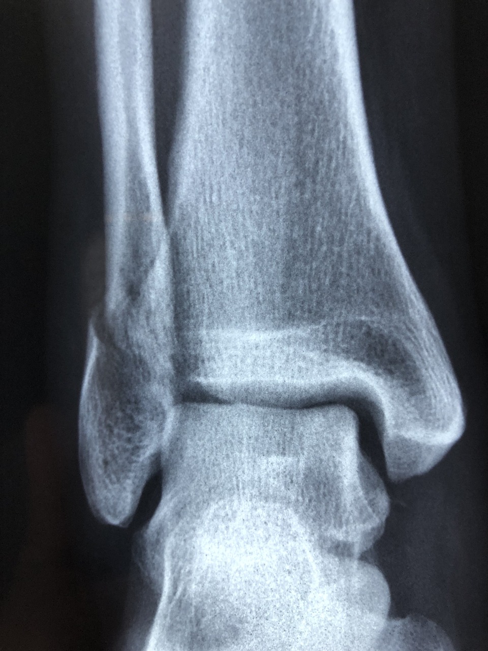
Diagnosing fibrocartilaginous mesenchymoma of bone requires a combination of imaging studies. X-rays, CT scans, and MRI scans are commonly used to assess the tumor's size, location, and impact on surrounding structures. Each imaging method provides unique insights:
X-rays reveal osteolytic areas with a lobulated framework, often showing radiolucent and radiodense foci.
CT scans help identify cortical destruction and provide detailed images of the bone.
MRI scans highlight cartilaginous calcifications and show strong contrast uptake, indicating the tumor's vascularity and aggressive potential.
These imaging techniques play a crucial role in visualizing the tumor and guiding further diagnostic steps.
Role of Biopsy in Confirming Diagnosis
A biopsy is essential for confirming the diagnosis of fibrocartilaginous mesenchymoma of bone. It involves analyzing a tissue sample under a microscope to identify the tumor's unique characteristics. In some cases, initial biopsy results may lead to misdiagnosis. For example:
A low-grade spindle cell lesion without osteoid or chondroid matrix may resemble desmoplastic fibroma.
Negative immunostaining for specific markers helps rule out other conditions.
Thorough examination of the excised mass often reveals features consistent with fibrocartilaginous mesenchymoma, such as cartilage nodules and fibrous stroma. Accurate biopsy interpretation is critical for distinguishing this tumor from other bone lesions.
Differentiating from Other Bone Tumors
Fibrocartilaginous mesenchymoma of bone shares similarities with other bone tumors, making differentiation challenging. Common differential diagnoses include fibrous dysplasia, desmoplastic fibroma, low-grade fibrosarcoma, chondromyxoid fibroma, and low-grade chondrosarcoma. However, histological analysis provides key distinctions. The tumor's unique combination of epiphyseal plate-like cartilage and dense fibrous stroma sets it apart.
In some cases, small biopsy samples may lack cartilaginous nodules, leading to misdiagnosis. For instance, a lytic lesion in a 36-year-old male was initially identified as desmoplastic fibroma. Further examination of the excised mass confirmed fibrocartilaginous mesenchymoma. This highlights the importance of comprehensive histological evaluation in ensuring an accurate diagnosis.
Treatment Options for Fibrocartilaginous Mesenchymoma of Bone
Surgical Management of the Tumor
Surgical resection serves as the primary treatment for fibrocartilaginous mesenchymoma of bone. This procedure involves removing the tumor along with any surrounding tissue that may contain cancerous cells. The surgical approach depends on the tumor's size and location. Smaller tumors may be treated using minimally invasive techniques, such as arthroscopy, which requires only small incisions. Larger tumors often necessitate open surgery, involving a more extensive incision to ensure complete removal. In some cases, surgeons may need to perform a bone graft to replace the removed segment and restore structural integrity. Early surgical intervention is critical to prevent complications and preserve the function of the affected bone.
Chemotherapy and Radiation Therapy
Chemotherapy is rarely used for fibrocartilaginous mesenchymoma of bone due to the tumor's resistance to this treatment. However, radiation therapy is a more common option, particularly when surgery is not feasible. Radiation therapy employs high-energy radiation to destroy cancerous cells and inhibit their growth. It is often used as an alternative treatment when surgical resection is not possible or when the tumor is located in a challenging area. In rare cases where the tumor has metastasized, chemotherapy may be considered, but its effectiveness remains limited. Radiation therapy plays a vital role in managing this condition when surgical options are restricted.
Post-Treatment Rehabilitation and Care
Post-treatment rehabilitation focuses on restoring normal function to the affected area. Patients often undergo physical therapy to regain mobility and strength in the impacted limb. Rehabilitation programs aim to enhance movement capabilities and improve the patient's overall quality of life. Regular imaging tests are also essential to monitor for any signs of tumor recurrence. These follow-up measures ensure that any potential issues are detected early, allowing for prompt intervention. Comprehensive rehabilitation and monitoring are crucial for achieving the best possible outcomes after treatment.
Prognosis and Follow-Up Care
Long-Term Outcomes for Patients
Patients diagnosed with fibrocartilaginous mesenchymoma of bone generally experience favorable long-term outcomes. This rare tumor has a high survival rate when treated appropriately. Complete surgical resection significantly reduces the risk of recurrence, allowing patients to live healthy lives. Studies show that local recurrences are rare when the tumor is entirely removed. With proper care and monitoring, most individuals regain full functionality in the affected area and maintain a good quality of life.
Risk of Recurrence and Monitoring
The recurrence rate for fibrocartilaginous mesenchymoma of bone remains low, especially after complete surgical removal. In rare cases, recurrence has occurred when the tumor was not fully excised. For instance, one patient experienced a recurrence within a year following an incomplete resection. To minimize this risk, regular follow-up imaging is essential. Common imaging modalities used for monitoring include:
X-rays for assessing bone structure.
CT scans for detailed cross-sectional images.
MRI scans for evaluating soft tissue involvement.
These tools help detect any signs of recurrence early, ensuring timely intervention.
Importance of Regular Follow-Up
Regular follow-up care plays a critical role in managing fibrocartilaginous mesenchymoma of bone. Patients should adhere to scheduled imaging tests and clinical evaluations to monitor for potential recurrence. Physical therapy may also be necessary to restore mobility and strength in the affected limb. Maintaining follow-up appointments ensures that any complications are addressed promptly. Early detection of recurrence or other issues can significantly improve long-term outcomes. Consistent monitoring and rehabilitation contribute to a better prognosis and overall well-being.
Fibrocartilaginous mesenchymoma of bone is a rare tumor with unique histological features and a favorable prognosis when treated effectively. Complete surgical resection remains the cornerstone of treatment, as it significantly reduces the risk of local recurrence. Unlike other bone tumors, this condition shows no genetic link to fibrous dysplasia or low-grade osteosarcoma, as recent molecular studies have confirmed.
Early diagnosis plays a critical role in improving outcomes. Timely intervention allows for better tumor management, minimizes complications, and enhances the patient’s quality of life. Clinicians should consider this tumor in cases of unexplained bone pain, swelling, or fractures.
Ongoing research continues to explore innovative treatment approaches. For example, targeted therapies like Drug A and Drug B show promise in shrinking tumor size and preventing metastasis:
Drug Name | Description | Early Results |
|---|---|---|
Drug A | A targeted therapy inhibiting a specific signaling pathway involved in tumor growth. | Promising results in shrinking tumor size and improving survival rates. |
Drug B | An experimental drug targeting a different molecular pathway associated with the tumor. | Preliminary data suggests effectiveness in inhibiting growth and preventing metastasis. |
These advancements offer hope for more effective and less invasive treatment options in the future. Early detection, multidisciplinary care, and ongoing research remain vital in ensuring the best outcomes for patients with fibrocartilaginous mesenchymoma of bone.
FAQ
What makes fibrocartilaginous mesenchymoma of bone unique?
Fibrocartilaginous mesenchymoma combines spindle cells, cartilage nodules, and bone trabeculae. Its histological features mimic growth plate cartilage, which distinguishes it from other bone tumors. This rare tumor also shows locally aggressive behavior, requiring early diagnosis and treatment.
Can fibrocartilaginous mesenchymoma spread to other parts of the body?
This tumor rarely metastasizes. It primarily remains localized to the bone where it develops. However, its aggressive growth can damage surrounding tissues, emphasizing the importance of timely surgical intervention.
How is fibrocartilaginous mesenchymoma diagnosed?
Doctors use imaging techniques like X-rays, CT scans, and MRIs to assess the tumor. A biopsy confirms the diagnosis by identifying its unique histological features, such as cartilage nodules and spindle cells.
Is chemotherapy effective for treating this tumor?
Chemotherapy is not typically effective for fibrocartilaginous mesenchymoma. Surgical resection remains the primary treatment. Radiation therapy may be considered in cases where surgery is not feasible.
What is the prognosis for patients with fibrocartilaginous mesenchymoma?
The prognosis is excellent when the tumor is completely removed. Most patients recover fully and experience minimal risk of recurrence. Regular follow-up care ensures early detection of any potential issues.
---
ℹ️ Explore more: Read our Comprehensive Guide to All Known Cancer Types for symptoms, causes, and treatments.
See Also
Understanding Angiosarcoma: Key Information You Should Know
Chondrosarcoma: Symptoms and Essential Information Explained
Choriocarcinoma Overview: Treatment Options and Insights
All You Need to Know About Carcinoid Tumors
Essential Facts About Embryonal Carcinoma You Should Understand

© 2026 Banish Cancer. Banish Cancer is a registered non-profit organization providing evidence-based research, educational courses, and Fear Response AI support services. Reg. No: 305706884 | Address: Taikos pr. 43-603, Kaunas, Lithuania.