Ganglioneuroma Explained: Key Facts You Should Know

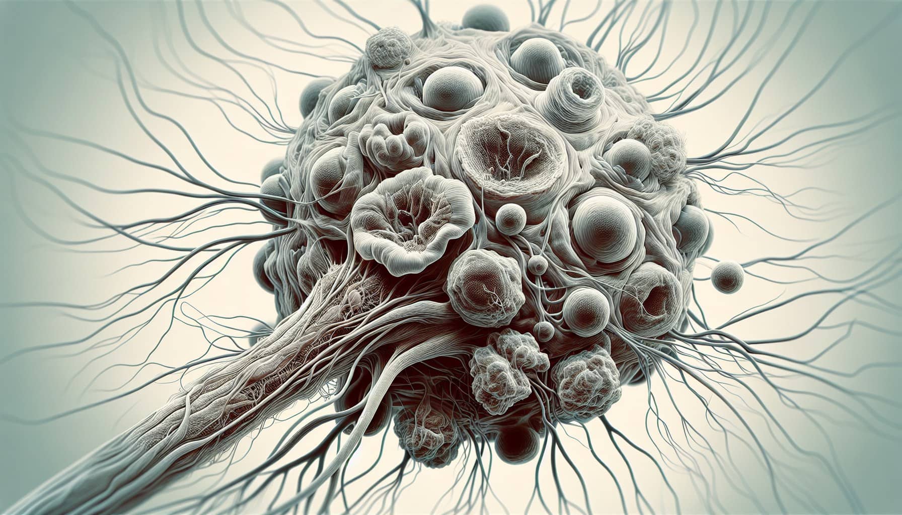
Ganglioneuroma is a rare, benign tumor that develops from autonomic nerve cells. It originates from neural crest sympathogonia cells, which are part of the sympathetic nervous system. These tumors are fully mature and lack immature elements, reflecting their differentiated nature. You typically find ganglioneuromas in individuals over 10 years old. Although they grow slowly and remain non-cancerous, their location in areas like the adrenal glands or chest can sometimes cause symptoms. Most cases are discovered incidentally during imaging for unrelated conditions, emphasizing their often silent presence.
Key Takeaways
Ganglioneuroma is a rare, non-cancerous tumor that grows slowly. It often shows no symptoms and is found by accident during scans for other problems.
These tumors usually grow in the adrenal glands, chest, belly, or neck. Their location might cause discomfort or other issues sometimes.
Surgery is the main way to treat ganglioneuromas with symptoms. Watching and checking regularly is an option for tumors without symptoms.
The outlook for ganglioneuroma is very good. After full surgery, it rarely comes back. Most people recover completely with good care.
If you have strange symptoms like lasting pain or trouble breathing, see a doctor quickly for early help and treatment.
Characteristics of Ganglioneuroma

Benign Nature
Ganglioneuroma is a non-cancerous tumor that grows slowly over time. Its benign nature means it rarely transforms into a malignant form. You can feel reassured knowing that these tumors typically have well-defined borders and do not invade nearby tissues or organs. Most cases remain asymptomatic, and doctors often discover them incidentally during imaging studies for unrelated issues.
🧠 Did you know? Ganglioneuromas are composed of mature cells, which explains their slow growth and lack of aggressive behavior.
Typical Locations
Ganglioneuromas commonly develop in areas associated with the sympathetic nervous system. These include the adrenal glands, chest (posterior mediastinum), abdomen (retroperitoneum), and neck. Among these, the posterior mediastinum accounts for about 41% of cases, while the retroperitoneum represents 37%. Less frequently, these tumors appear in the head, pelvis, or even the gastrointestinal tract.
Most common locations:
Posterior mediastinum
Retroperitoneum
Adrenal glands
Neck
The sympathetic nervous system plays a key role in their development. Ganglioneuromas arise from neural crest sympathogonia, which are cells that mature into ganglion cells and nerve fibers. This connection explains why these tumors are often found along the pathways of the sympathetic nerves.
Cellular Composition
The cellular makeup of ganglioneuroma sets it apart from malignant tumors. It consists of mature ganglion cells, Schwann cells, and nerve fibers. These components contribute to its benign behavior. Unlike malignant tumors such as neuroblastoma, ganglioneuroma does not invade blood vessels or nearby organs. Its fully differentiated nature ensures it lacks immature or aggressive elements.
Key takeaway: The absence of immature cells in ganglioneuroma highlights its non-cancerous and stable characteristics.
Doctors often rely on imaging studies to identify these tumors. Homogeneous and well-defined masses on scans further confirm their benign nature.
Symptoms and Signs
Asymptomatic Cases
Ganglioneuroma often presents without any noticeable symptoms. You may not even realize its presence until it is discovered during imaging tests for unrelated health concerns. These tumors typically grow slowly and remain painless, which makes them hard to detect without medical evaluation.
Note: Most ganglioneuromas are benign and asymptomatic, making incidental discovery during routine check-ups or imaging studies quite common.
Common Symptoms
When symptoms do occur, they usually depend on the tumor's size and location. Larger tumors can press against nearby organs or structures, leading to discomfort or other issues.
Pain or discomfort: You might experience pain in the area where the tumor is located. For example, tumors in the abdomen can cause abdominal pain, while those near the spine may lead to back pain.
Pressure on nearby organs: Tumors in the chest can compress the windpipe, causing difficulty breathing or chest pain. Similarly, abdominal tumors may lead to bloating or a feeling of fullness.
Hormonal imbalances: If the tumor affects the adrenal glands, it can disrupt hormone production. This may result in symptoms like fatigue, high blood pressure, or other hormonal changes.
In rare cases, tumors near the spinal cord can cause spinal deformities or nerve compression. This might lead to pain, weakness, or loss of sensation in your limbs.
Tip: If you notice persistent pain, breathing difficulties, or unusual symptoms, consult a healthcare provider. Early diagnosis can help manage potential complications effectively.
Causes and Risk Factors
Genetic Factors
Ganglioneuroma has been linked to specific genetic mutations and syndromes. One notable connection is with Cowden syndrome, which involves a mutation in the PTEN gene. This mutation can disrupt normal cell growth and division, potentially contributing to tumor development. Another association exists with diffuse ganglioneuromatosis, a condition tied to multiple endocrine neoplasia type IIB (MENIIB). Neurofibromatosis type 1 (NF1), a genetic disorder that causes tumors to form on nerve tissue, has also been identified as a less common contributor. If you or a family member has a history of these conditions, it may increase the likelihood of developing ganglioneuroma.
Tip: If you suspect a genetic predisposition, consider consulting a healthcare provider for genetic testing and early detection strategies.
Developmental Origins
Ganglioneuroma originates from neural crest cells, which play a vital role in the development of your nervous system. These cells give rise to sympathetic ganglion cells, the primary components of ganglioneuroma. This tumor represents a mature stage of peripheral neuroblastic tumors, meaning it has fully differentiated and lacks immature elements.
Unlike many other health conditions, ganglioneuroma rarely results from environmental or lifestyle factors. Its developmental origins are rooted in the early stages of your body's formation, making it a condition that is largely beyond your control. Understanding this can help you focus on monitoring and managing symptoms rather than worrying about preventable causes.
Key takeaway: Ganglioneuroma develops from neural crest tissue during early development, emphasizing its genetic and biological roots over external influences.
Diagnosis of Ganglioneuroma
Imaging Studies
Doctors often use imaging studies to identify and evaluate ganglioneuroma. These tests help determine the tumor's size, location, and impact on surrounding tissues.
MRI and CT Scans:
MRI and CT scans are the most effective tools for diagnosing ganglioneuroma. CT scans provide detailed images of the tumor's size, origin, and any involvement with nearby structures. MRI offers better tissue contrast, making it ideal for assessing the tumor's relationship with surrounding organs.
Here’s a quick comparison of imaging techniques:
Imaging Technique | Effectiveness | Limitations |
|---|---|---|
CT Scan | Superior for identifying tumor size, organ of origin, tissue invasion, vascular encasement, adenopathy, and calcifications. | Standard scans may miss small lesions or subtle invasions. |
MRI | Better tissue discrimination, effective for evaluating organs of origin and regional invasion. | More expensive and less available than CT. |
MIBG Scintigraphy | High sensitivity (88%) and specificity (99%) for sympathetic tissue tumors. | Only 70% of ganglioneuromas take up MIBG; cannot distinguish tumor types. |
Ultrasound:
In some cases, doctors may use ultrasound as an initial evaluation tool. This method is non-invasive and can provide a quick overview of the tumor's location and size. However, it is less detailed than MRI or CT scans.
Biopsy
A biopsy is essential for confirming the diagnosis of ganglioneuroma. During this procedure, a small tissue sample is taken from the tumor and analyzed under a microscope.
Tissue Analysis:
The biopsy helps identify the tumor's cellular composition, confirming its benign nature. It also ensures that the tumor is not a more aggressive type, such as neuroblastoma.Differentiation:
This step is crucial for distinguishing ganglioneuroma from other nerve-related tumors. Accurate diagnosis allows doctors to plan the most effective treatment.
Additional Tests
Doctors may recommend additional tests to gather more information about the tumor and its effects on your body.
Blood Tests:
Blood tests can detect hormonal imbalances, especially if the tumor involves the adrenal glands. These imbalances may cause symptoms like high blood pressure or fatigue.Genetic Testing:
If you have a family history of genetic conditions like neurofibromatosis type 1, genetic testing may be suggested. This can help identify hereditary factors that might contribute to the tumor's development.
Tip: Early and accurate diagnosis ensures better management of ganglioneuroma. If you experience symptoms or have concerns, consult your healthcare provider promptly.
Treatment and Prognosis
Surgical Removal
Surgical removal is the primary treatment for ganglioneuroma when the tumor causes symptoms or grows significantly. Doctors recommend surgery if the tumor exerts pressure on nearby organs, leading to discomfort or functional issues. For instance, tumors pressing on the spinal cord may cause nerve compression or even paralysis. Surgery is also necessary for long-standing tumors that have caused irreversible damage.
When is surgery needed?
The tumor causes symptoms like pain or hormonal imbalances.
It exerts pressure on critical structures, such as the spinal cord or nearby organs.
It leads to physical deformities or functional impairments.
Minimally invasive techniques, such as laparoscopic surgery, may be an option depending on the tumor's size and location. These methods reduce recovery time and minimize risks.
Tip: Early surgical intervention can prevent complications and improve outcomes.
Observation
Not all ganglioneuromas require immediate treatment. If the tumor is asymptomatic and stable, doctors may recommend regular monitoring instead of surgery. This approach avoids unnecessary risks associated with invasive procedures.
During observation, you may undergo periodic imaging studies to track the tumor's growth. If the tumor remains unchanged and does not cause symptoms, no further action is needed. However, any signs of growth or new symptoms would prompt a reevaluation of treatment options.
Note: Observation is a safe and effective strategy for managing benign and asymptomatic tumors.
Prognosis
The prognosis for ganglioneuroma is excellent. After complete surgical removal, recurrence rates are extremely low. Studies show no recurrence or metastasis in patients who underwent successful tumor resection. This highlights the importance of complete removal during surgery.
Complications are rare but may occur if the tumor affects critical structures. For example, untreated tumors pressing on the spinal cord can lead to paralysis. Hormonal imbalances caused by adrenal gland involvement may result in high blood pressure or severe diarrhea. Despite these risks, most patients recover fully after treatment.
Key takeaway: With proper management, ganglioneuroma has a favorable outcome and minimal risk of recurrence.
Ganglioneuroma is a rare, benign tumor that typically grows slowly and remains asymptomatic. It may cause symptoms like pain, bloating, or difficulty breathing if it presses on nearby organs. Surgical removal is the primary treatment for symptomatic cases, and the prognosis is highly favorable with proper care. Most cases are manageable, and early diagnosis ensures better outcomes.
Tip: If you notice unusual symptoms or have concerns, consult a healthcare provider promptly. Early evaluation can help address potential issues effectively.
FAQ
What is the difference between ganglioneuroma and neuroblastoma?
Ganglioneuroma is a benign tumor made of mature nerve cells, while neuroblastoma is a cancerous tumor with immature cells. Ganglioneuroma grows slowly and doesn’t spread, but neuroblastoma can grow aggressively and metastasize.
Tip: Early diagnosis helps differentiate these tumors and ensures proper treatment.
Can ganglioneuroma turn into cancer?
No, ganglioneuroma rarely becomes cancerous. Its mature cells make it a stable, non-aggressive tumor. Regular monitoring ensures no unexpected changes occur.
🧠 Did you know? Ganglioneuroma is one of the most benign nerve-related tumors.
How is ganglioneuroma treated?
Doctors usually recommend surgery for symptomatic or growing tumors. Asymptomatic cases may only need observation with regular imaging to track growth.
Treatment options:
Surgical removal
Observation for stable tumors
Is ganglioneuroma hereditary?
Ganglioneuroma may have genetic links, especially with conditions like neurofibromatosis type 1. However, most cases occur sporadically without a family history.
Note: Genetic testing can help identify hereditary risks.
Can ganglioneuroma cause hormonal problems?
Yes, if the tumor affects the adrenal glands, it may disrupt hormone production. This can lead to symptoms like high blood pressure, fatigue, or other imbalances.
Key takeaway: Hormonal symptoms often resolve after tumor removal.
---
ℹ️ Explore more: Read our Comprehensive Guide to All Known Cancer Types for symptoms, causes, and treatments.
See Also
Essential Insights Into Embryonal Carcinoma You Should Know
Craniopharyngioma: Important Features You Need To Understand
Key Information About Carcinoid Tumors You Must Know

© 2026 Banish Cancer. Banish Cancer is a registered non-profit organization providing evidence-based research, educational courses, and Fear Response AI support services. Reg. No: 305706884 | Address: Taikos pr. 43-603, Kaunas, Lithuania.