What Is a Gastrointestinal Carcinoid Tumor?

A gastrointestinal carcinoid tumor is a rare type of cancer that grows slowly in your digestive tract. It develops from neuroendocrine cells, which produce hormones that help regulate digestion. These cells control the release of digestive juices and the movement of food through your stomach and intestines.
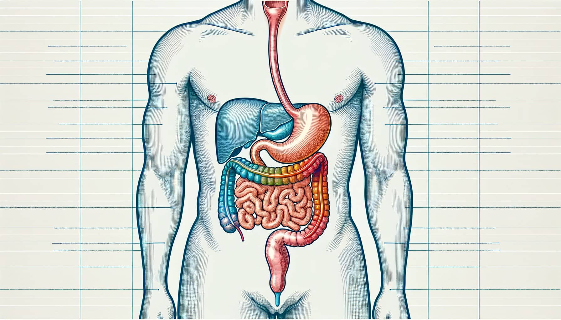
This tumor often appears in specific areas of the gastrointestinal tract. The appendix is the most common site, accounting for 35% of cases. The ileum, part of the small intestine, follows at 28%, while the rectum accounts for 13%.
Understanding where these tumors form and their connection to hormone-secreting cells can help you recognize their significance in digestive health.
Key Takeaways
Gastrointestinal carcinoid tumors are rare cancers in the digestive system. They start in special cells called neuroendocrine cells. Finding them early can lead to better treatment results.
Common signs include red, warm skin on the face, bad diarrhea, and trouble breathing. See a doctor if symptoms last or seem strange.
Surgery is usually the first choice to treat small tumors. Medicines like somatostatin analogs can control symptoms and slow tumor growth.
Regular check-ups are important to watch your health. They help find new tumors early and manage leftover symptoms.
Support groups and counseling can give emotional and practical help. Talking to others can make living with this condition easier.
Characteristics and Development

What is a gastrointestinal carcinoid tumor made of?
A gastrointestinal carcinoid tumor forms from neuroendocrine cells in your digestive tract. These cells originate from primitive stem cells and play a role in hormone production. Tumors often develop deep within the mucosa, creating small, firm nodules that protrude into the intestinal lumen.
Histologically, these tumors display five distinct patterns: solid, trabecular, tubules and glands, poorly differentiated, and mixed patterns. Midgut carcinoid tumors, found in the small intestine, are argentaffin positive and produce high levels of serotonin and other vasoactive peptides. In contrast, hindgut carcinoid tumors, located in the rectum, are argentaffin negative and rarely secrete these substances.
Common locations in the gastrointestinal tract
Neuroendocrine cells are scattered throughout your digestive system, but certain areas are more prone to tumor development. These include the small intestine, appendix, and rectum.
Location | Susceptibility Reason |
|---|---|
Small Intestine | Contains a high density of neuroendocrine cells that can develop into tumors. |
Appendix | Similar to the small intestine, it has a concentration of neuroendocrine cells. |
Rectum | Also has neuroendocrine cells, contributing to tumor formation. |
The presence of neuroendocrine cells in these regions increases the likelihood of a gastrointestinal carcinoid tumor forming.
How gastrointestinal carcinoid tumors grow and spread
The growth and spread of gastrointestinal carcinoid tumors depend on their grade. Tumors are classified into three grades based on their growth rate and cellular characteristics:
Grade | Description | Growth Rate |
|---|---|---|
1 | Low grade | Slow |
2 | Intermediate | Moderate |
3 | High grade | Fast |
Grade 1 and Grade 2 tumors grow slowly but may spread to other parts of your body over time. Grade 3 tumors, however, grow rapidly and are more likely to metastasize. Understanding these growth patterns can help you recognize the importance of early diagnosis and treatment.
Symptoms and Signs
Common symptoms of gastrointestinal carcinoid tumors
Gastrointestinal carcinoid tumors can cause a variety of symptoms, many of which result from the hormones these tumors release. You might experience:
Facial flushing, which feels like warmth and redness spreading across your face.
Severe diarrhea that disrupts your daily life.
Wheezing or difficulty breathing.
A fast heartbeat, even when resting.
Muscle weakness or fatigue.
Pain, nausea, or loss of appetite.
Symptoms of anemia, such as feeling tired or short of breath.
In some cases, you may notice changes in your skin, like stretch marks or increased body and facial hair. These symptoms often depend on the tumor's activity and the hormones it produces.
How symptoms vary by tumor location
The location of the tumor in your gastrointestinal tract plays a significant role in the symptoms you experience. The table below highlights how symptoms differ based on where the tumor forms:
Location | Symptoms |
|---|---|
Appendix | Often asymptomatic; may cause appendicitis with fever, nausea, vomiting, and abdominal pain. |
Small Intestine | Can cause cramps, belly pain, weight loss, fatigue, bloating, diarrhea, and nausea/vomiting. |
Colon | Similar to small intestine; may lead to intestinal bleeding and anemia. |
Rectum | Pain, bleeding, and constipation; often found during routine exams. |
Stomach | Usually asymptomatic; may cause carcinoid syndrome symptoms. |
Understanding these variations can help you identify potential warning signs based on your symptoms.
When to seek medical attention
You should consult a healthcare provider if you notice persistent or unusual symptoms. Warning signs that require immediate attention include:
Severe diarrhea or facial flushing.
A fast heartbeat or wheezing.
Black, tarry stools, which may indicate bleeding.
Persistent pain, nausea, or loss of appetite.
Symptoms of anemia, such as fatigue or shortness of breath.
Early diagnosis of a gastrointestinal carcinoid tumor can improve treatment outcomes. If you experience any of these symptoms, don't hesitate to seek medical advice.
Diagnosis
Medical history and physical examination
Diagnosing a gastrointestinal carcinoid tumor often begins with your medical history and a physical exam. Your doctor may ask about your symptoms, lifestyle, and any risk factors, such as a family history of neuroendocrine tumors. They will also inquire about signs of carcinoid syndrome, like facial flushing or diarrhea. During the physical exam, they may check for abdominal tenderness, masses, or other abnormalities that could indicate a tumor.
Diagnostic tests and imaging techniques
Doctors use several diagnostic tests to identify gastrointestinal carcinoid tumors. These tests help visualize the tumor, assess its spread, and confirm its presence. Below is a table summarizing the most common diagnostic methods:
Diagnostic Test | Description |
|---|---|
Questions about health, lifestyle, symptoms, and risk factors, including those related to carcinoid syndrome. | |
Imaging tests | Includes barium x-ray and CT scans to visualize abnormalities and check for tumor spread. |
Blood and urine tests | Helpful in diagnosing carcinoid syndrome in symptomatic patients. |
Imaging techniques like CT and MRI scans play a crucial role in detecting and monitoring these tumors. CT scans are particularly effective for identifying liver metastases and mesenteric masses. MRI scans can sometimes detect liver metastases more effectively than CT scans. Other imaging methods, such as radionuclide scans and octreoscans, help locate tumors and evaluate treatment effectiveness. However, small-bowel carcinoids can be challenging to detect, highlighting the limitations of these techniques.
Biopsy and pathology for confirmation
A biopsy is essential for confirming the diagnosis of a gastrointestinal carcinoid tumor. During this procedure, your doctor removes a small tissue sample from the suspected tumor. Pathologists then examine the sample under a microscope to check for cancerous cells. If the biopsy does not provide enough tissue, surgical intervention may be necessary to obtain a more substantial sample. This step is critical for determining the tumor type and guiding your treatment plan.
Treatment Options

Surgical removal of the tumor
Surgery is often the first step in treating a gastrointestinal carcinoid tumor, especially when the tumor is localized. The type of surgery depends on the tumor's size and location. For small tumors on the lining of your gastrointestinal tract, doctors may perform an endoscopic resection. This minimally invasive procedure removes the tumor without major surgery. If the tumor is larger than 2 cm, a more extensive surgery, such as a resection, may be necessary. This involves removing part of the affected organ, such as the rectum or duodenum, along with nearby lymph nodes.
In some cases, surgeons may perform a pancreatoduodenectomy for larger tumors in the duodenum. This procedure removes part of the pancreas, duodenum, and surrounding tissues. These surgical options aim to eliminate the tumor and reduce the risk of recurrence.
Medications and hormone therapy
Medications play a crucial role in managing symptoms and slowing tumor growth. Somatostatin analogs, such as octreotide and lanreotide, are commonly prescribed. These drugs help control symptoms like diarrhea and flushing while also inhibiting tumor growth. Octreotide is often used to treat carcinoid syndrome and may temporarily shrink tumors. Lanreotide, administered monthly, offers similar benefits.
For severe diarrhea, doctors may recommend telotristat (Xermelo™) alongside somatostatin analogs. Advanced tumors may require targeted drugs like everolimus (Afinitor®), which can slow tumor progression. In some cases, interferon-alfa is used to improve symptoms and reduce tumor growth. These therapies provide effective options for managing both localized and metastatic tumors.
Additional treatments, such as radiation or chemotherapy
Radiation and chemotherapy are typically reserved for specific situations. Radiation therapy helps patients who cannot undergo surgery or those with residual tumors after surgery. It can also relieve symptoms when the cancer has spread to other parts of the body.
Chemotherapy is usually recommended for metastatic carcinoid tumors, especially when other treatments fail or the tumors grow rapidly. It may also be used to manage severe symptoms caused by the tumor. These treatments, while not always the first choice, offer valuable options for advanced cases.
Note: Recent advancements in treatment focus on personalized approaches. Clinical trials are exploring new therapies, including targeted treatments, to improve outcomes for patients with gastrointestinal carcinoid tumors.
Prognosis and Living with a Gastrointestinal Carcinoid Tumor
Factors influencing prognosis
Several factors can influence your prognosis if you have a gastrointestinal carcinoid tumor. These include the tumor's location, symptoms, and stage at diagnosis. The table below highlights key factors and their impact on survival:
Factor | Influence on Prognosis |
|---|---|
Site of Origin | Significant; foregut and midgut have lower survival than hindgut |
Symptomatic Presentation | Strongly affects survival; foregut carcinoids always symptomatic, midgut 61%, hindgut 37% (p < 0.001) |
Size of Primary Tumor | Not an independent predictor of survival |
Presence of Liver Metastases | Not an independent predictor of survival |
Localized tumors tend to have better outcomes. For example, the five-year survival rate for localized tumors is 97%, while distant-stage tumors have a survival rate of 68%. Early detection plays a crucial role in improving your prognosis.
Long-term management and follow-up care
Managing a gastrointestinal carcinoid tumor requires a combination of treatments and regular follow-ups. Surgical removal of the tumor and affected lymph nodes offers the best chance of a cure. If surgery isn't possible, treatments like somatostatin drugs can help control symptoms and slow tumor growth.
Follow-up care depends on the tumor's size and grade. Small, low-grade tumors may require minimal follow-up, while larger or higher-grade tumors need more frequent monitoring. For example:
Tumors smaller than 1 cm may need little to no follow-up.
Larger tumors or those with lymph node involvement may require yearly CT scans for three years, then every 1-2 years for the next seven years.
Blood and urine tests, such as 5-HIAA and Chromogranin A, can help monitor your condition but aren't always necessary.
Regular follow-ups help detect recurrence early and manage any lingering symptoms effectively.
Coping strategies and support resources
Living with a gastrointestinal carcinoid tumor can feel overwhelming, but you don't have to face it alone. Many resources are available to support you emotionally and practically. These include:
Online support groups and survivor networks.
Free rides and lodging during treatment.
Peer-matching services and 24/7 cancer helplines.
Organizations like CancerCare and the Carcinoid Cancer Foundation offer online forums, educational materials, and resource referrals. Connecting with others who share similar experiences can provide comfort and valuable insights.
Tip: Joining a support group or speaking with a counselor can help you manage stress and maintain a positive outlook during your treatment journey.
Understanding a gastrointestinal carcinoid tumor involves recognizing its characteristics, symptoms, and treatment options. These tumors often develop in the small intestine, rectum, or appendix and grow slowly, making early detection crucial. Below are some key takeaways:
Key Takeaway | Details |
|---|---|
Annual Diagnoses | Approximately 8,000 carcinoid tumors are diagnosed each year in the U.S. |
Common Locations | Small intestine, rectum, colon, appendix, and stomach are the most common sites. |
Demographics | Average age of diagnosis is early 60s; more common in African Americans and slightly more in women than men. |
Increasing Incidence | The number of diagnosed cases has been rising, possibly due to improved detection methods. |
Early diagnosis and treatment significantly improve outcomes. Studies show that patients diagnosed early, especially those without advanced symptoms, experience better survival rates and quality of life. Aggressive therapies, including surgery and medications, can effectively manage even advanced cases.
If you notice unusual symptoms or have concerns, consult a healthcare professional. Personalized advice and timely care can make a significant difference in managing this condition.
FAQ
What causes gastrointestinal carcinoid tumors?
Doctors don't know the exact cause. These tumors form when neuroendocrine cells in your digestive tract grow uncontrollably. Genetic mutations may play a role. Some people inherit conditions like multiple endocrine neoplasia type 1 (MEN1), which increases their risk.
Tip: If you have a family history of neuroendocrine tumors, consider genetic counseling.
Are gastrointestinal carcinoid tumors always cancerous?
No, not all carcinoid tumors are cancerous. Some remain benign and grow slowly without spreading. However, others can become malignant and metastasize to other organs. Early detection helps determine the tumor's nature and guides treatment.
Can gastrointestinal carcinoid tumors be prevented?
You can't completely prevent these tumors. However, maintaining a healthy lifestyle and addressing risk factors like smoking may reduce your chances. Regular check-ups and screenings help detect tumors early, especially if you have a family history.
How long does treatment for a gastrointestinal carcinoid tumor take?
Treatment duration depends on the tumor's size, location, and stage. Surgery may require a few weeks of recovery. Hormone therapy or medications could last months or years. Your doctor will create a personalized plan based on your condition.
Do gastrointestinal carcinoid tumors return after treatment?
Yes, these tumors can return, especially if they were advanced or not fully removed. Regular follow-ups, including imaging and blood tests, help monitor for recurrence. Early detection of returning tumors improves treatment outcomes.
Note: Discuss a follow-up schedule with your doctor to stay proactive about your health.
---
ℹ️ Explore more: Read our Comprehensive Guide to All Known Cancer Types for symptoms, causes, and treatments.
See Also
Essential Insights About Carcinoid Tumors You Need
Understanding Carcinoid Syndrome: Symptoms You Should Recognize
Choriocarcinoma Overview: Treatment Options and Information

© 2026 Banish Cancer. Banish Cancer is a registered non-profit organization providing evidence-based research, educational courses, and Fear Response AI support services. Reg. No: 305706884 | Address: Taikos pr. 43-603, Kaunas, Lithuania.