Molecular Features of Giant Cell Carcinoma of the Lung


Giant-cell carcinoma of the lung represents a rare and aggressive form of non-small cell lung cancer. It accounts for only 0.3% to 0.4% of NSCLC cases, with pure forms being even rarer. This subtype is associated with a poor prognosis, with five-year survival rates ranging from 10% to 11.8%. Its molecular profile, including unique genetic mutations and protein expression patterns, contributes to its aggressive behavior. These features not only distinguish it from other lung cancers but also highlight the need for advanced diagnostic and therapeutic approaches.
Key Takeaways
Giant-cell carcinoma of the lung is a rare, fast-growing cancer. It makes up only 0.3% to 0.4% of lung cancer cases. Finding it early can lead to better results.
Special gene changes, like TP53 and PIK3CA, affect how it grows. These changes also impact how the cancer reacts to treatments. Knowing about them helps pick the best therapy.
Biomarkers like cytokeratin (CK) and vimentin are very important. They help doctors confirm if it is giant-cell carcinoma. These markers also separate it from other lung cancers.
New tools, like liquid biopsy, check the cancer without surgery. They track how the cancer grows and responds to treatments, helping doctors care for patients better.
Targeted therapies and immunotherapy are hopeful new treatments. They focus on the tumor's unique traits to make treatments work better.
Introduction to Giant-Cell Carcinoma of the Lung
Defining Giant-Cell Carcinoma
Giant-cell carcinoma of the lung is a rare and aggressive variant of non-small cell lung cancer. It is characterized by unique histological features that set it apart from other lung cancer subtypes. These tumors consist of extremely large, bizarre, and highly pleomorphic cells. Malignant cells often exhibit multiple nuclei, with sizes averaging five times larger than lymphocyte nuclei. Significant variation in both cell and nuclear size is a hallmark of this carcinoma. The anaplastic nature of these cells reflects a lack of differentiation, and they typically appear as single cells or loosely cohesive aggregates. Hyperchromatic chromatin, prominent nucleoli, and multinucleated giant cells further define its morphology. These features contribute to the tumor's aggressive behavior and poor prognosis.
Epidemiology and Clinical Presentation
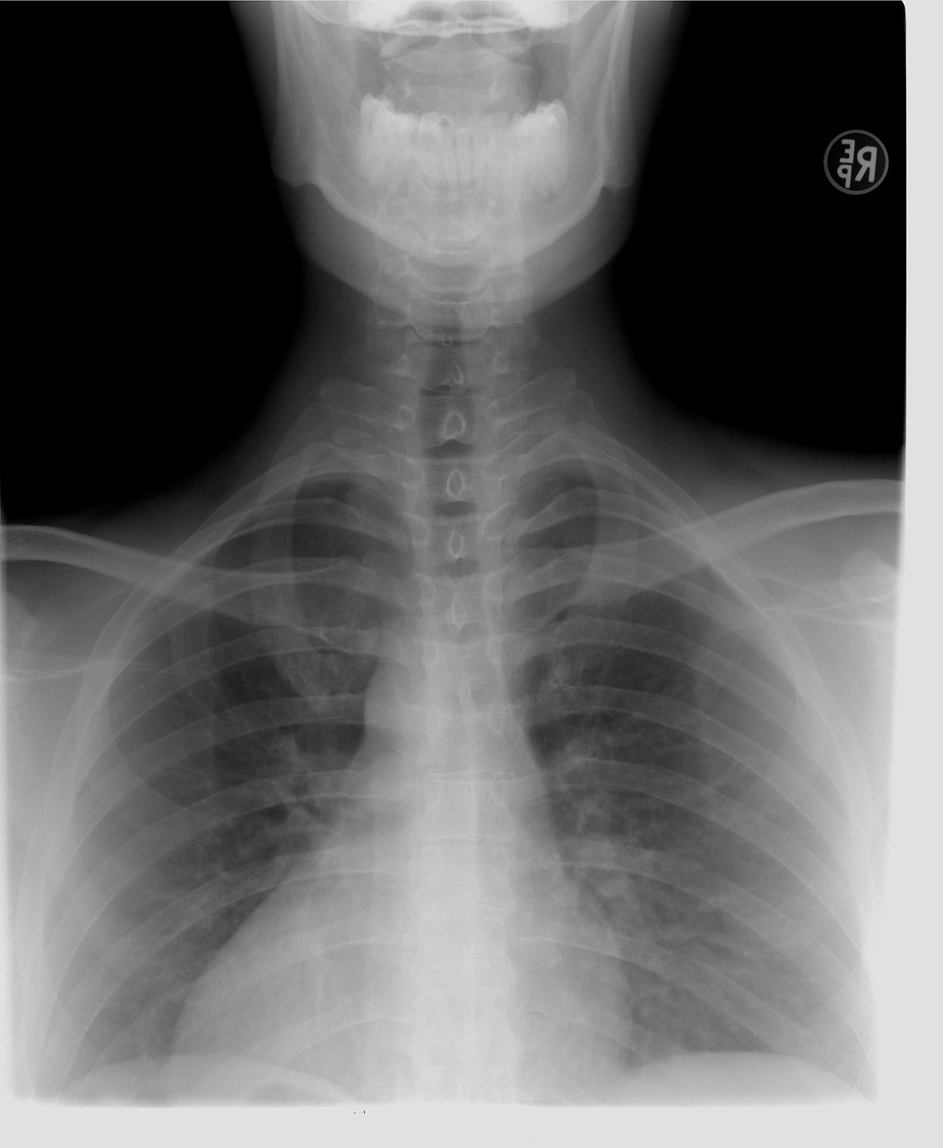
Giant-cell carcinoma of the lung is exceedingly rare, accounting for only 0.3% to 0.4% of primary pulmonary malignancies. In the United States, studies estimate an age-adjusted incidence rate of approximately three new cases per million individuals annually. This subtype predominantly affects older adults, with a higher prevalence in males. Patients often present with advanced-stage disease due to the tumor's rapid progression and aggressive nature. Common symptoms include persistent cough, chest pain, hemoptysis, and unexplained weight loss. These nonspecific symptoms frequently overlap with other lung cancer types, complicating early diagnosis. Imaging studies typically reveal large, irregular masses, but definitive diagnosis requires histopathological examination to identify the tumor's distinct cellular features.
Molecular Characteristics of Giant-Cell Carcinoma
Genetic Mutations
Giant-cell carcinoma of the lung exhibits a distinct genetic mutation profile that sets it apart from other non-small cell lung cancer subtypes. These mutations influence tumor behavior, progression, and treatment response. The table below highlights some of the most common genetic mutations identified in this rare carcinoma:
Genetic Mutation | Description |
|---|---|
EGFR | Hotspot mutation found in some samples |
EML4-ALK | Gene fusion identified in a sample with spindle and giant cells |
MET | Hotspot mutation detected |
BRAF | Hotspot mutation detected |
PIK3CA | Hotspot mutation detected |
TP53 | Hotspot mutation detected, also found in other cases |
KRAS | Missense mutation detected in some samples, but not in all |
KMT2D | Missense mutation detected |
SETD2 | Missense mutation detected |
TET2 | Missense mutation detected |
MAP3K | Missense mutation detected |
Unlike other NSCLC subtypes, giant-cell carcinoma of the lung often lacks KRAS mutations, which are commonly observed in adenocarcinomas. The presence of TP53 mutations in this carcinoma correlates with reduced chemotherapy sensitivity, further complicating treatment strategies.
Protein Expression Patterns
Protein expression patterns in giant-cell carcinoma of the lung reveal significant insights into its aggressive nature. Overexpression of proteins such as Ki-67, a marker of cellular proliferation, indicates rapid tumor growth. Additionally, aberrant expression of p53 protein, resulting from TP53 mutations, is frequently observed. This abnormality disrupts cell cycle regulation and contributes to the tumor's anaplastic characteristics. Immunohistochemical studies often detect high levels of vascular endothelial growth factor (VEGF), which promotes angiogenesis and supports tumor progression. These protein markers not only aid in understanding the tumor's biology but also serve as potential diagnostic and therapeutic targets.
Altered Signaling Pathways
Altered signaling pathways play a pivotal role in the pathogenesis of giant-cell carcinoma of the lung. Dysregulation of the PI3K/AKT/mTOR pathway, driven by mutations in PIK3CA, enhances cell survival and proliferation. Similarly, aberrations in the MAPK pathway, linked to mutations in BRAF and MAP3K, contribute to uncontrolled cell growth. The MET signaling pathway, often activated by MET mutations, promotes tumor invasiveness and metastasis. These alterations underscore the complexity of the molecular landscape in this carcinoma and highlight potential avenues for targeted therapies.
Understanding these molecular characteristics is essential for developing personalized treatment strategies and improving outcomes for patients with giant-cell carcinoma of the lung.
Diagnostic Implications of Molecular Features
Biomarker-Based Diagnostics
Biomarkers play a critical role in diagnosing giant-cell carcinoma of the lung. Immunohistochemical analysis has identified cytokeratin (CK) and vimentin as reliable biomarkers for this rare carcinoma. Tumor cells consistently exhibit positive reactivity to these markers, confirming their diagnostic value. In contrast, chromogranin A (CgA) and synaptophysin (Syn), commonly associated with neuroendocrine tumors, are negative in giant-cell carcinoma. This distinction helps differentiate it from other lung cancer subtypes. Biomarker-based diagnostics not only enhance accuracy but also reduce the likelihood of misdiagnosis, which is crucial given the aggressive nature of this carcinoma.
The identification of these biomarkers has also paved the way for personalized diagnostic approaches. For instance, CK and vimentin expression patterns can guide pathologists in confirming the diagnosis when histological features alone are inconclusive. This targeted approach ensures that patients receive timely and appropriate treatment.
Advances in Molecular Diagnostic Tools
Recent advancements in molecular diagnostic tools have revolutionized the detection of giant-cell carcinoma of the lung. Techniques such as next-generation sequencing (NGS) and polymerase chain reaction (PCR) enable the identification of genetic mutations and altered signaling pathways with high precision. These tools provide a comprehensive molecular profile of the tumor, which is essential for understanding its behavior and selecting effective therapies.
Liquid biopsy represents another significant innovation. This non-invasive method detects circulating tumor DNA (ctDNA) in the bloodstream, offering a real-time snapshot of the tumor's genetic landscape. Liquid biopsy is particularly valuable for monitoring disease progression and assessing treatment response. Additionally, immunohistochemistry (IHC) remains a cornerstone of molecular diagnostics. By detecting protein expression patterns, IHC complements genetic analyses and provides a holistic view of the tumor's molecular characteristics.
The integration of these advanced tools into clinical practice has improved diagnostic accuracy and expanded the possibilities for personalized medicine. As technology continues to evolve, these methods will likely become even more refined, further enhancing patient outcomes.
Pathological Insights into Giant-Cell Carcinoma

Histological and Molecular Correlation
Histological findings in giant-cell carcinoma of the lung often align closely with its molecular features, providing valuable insights into its pathology. Immunohistochemical analysis frequently reveals positive staining for cytokeratin (CK) and vimentin, which are markers of epithelial and mesenchymal origin, respectively. These findings correlate with specific genetic mutations that drive tumor behavior. For instance, KRAS mutations, such as p.G12D, are commonly associated with CK and vimentin positivity. Conversely, the absence of chromogranin A (CgA) and synaptophysin (Syn) immunostaining helps differentiate this carcinoma from neuroendocrine tumors. This distinction is further supported by the presence of TP53 mutations, such as p.R249K, which disrupt cell cycle regulation and contribute to the tumor's aggressive nature.
The table below summarizes the relationship between histological findings and molecular mutations in giant-cell carcinoma of the lung:
Histological Findings | Molecular Mutations |
|---|---|
Positive CK and vimentin immunostaining | KRAS mutation (p.G12D) |
Negative CgA and Syn immunostaining | TP53 mutation (p.R249K) |
Other mutations: KMT2D, SETD2, TET2, MAP3K |
This correlation between histological and molecular features underscores the importance of integrating both approaches for accurate diagnosis and understanding of tumor biology.
Tumor Microenvironment and Molecular Interactions
The tumor microenvironment plays a critical role in the progression and aggressiveness of giant-cell carcinoma of the lung. This carcinoma exhibits a highly dynamic microenvironment characterized by extensive angiogenesis, immune evasion, and stromal interactions. Overexpression of vascular endothelial growth factor (VEGF) promotes the formation of new blood vessels, ensuring a steady supply of nutrients to the rapidly growing tumor. Additionally, the presence of immunosuppressive molecules within the microenvironment hinders the activity of cytotoxic T cells, allowing the tumor to evade immune surveillance.
Molecular interactions within the microenvironment further enhance tumor progression. Dysregulated signaling pathways, such as PI3K/AKT/mTOR and MET, not only drive tumor cell proliferation but also influence the surrounding stromal cells. These interactions create a supportive niche that facilitates invasion and metastasis. Understanding these complex molecular dynamics is essential for developing therapies that target both the tumor and its microenvironment.
The interplay between histological findings, molecular features, and the tumor microenvironment highlights the multifaceted nature of giant-cell carcinoma of the lung. These insights pave the way for more effective diagnostic and therapeutic strategies.
Therapeutic Relevance of Molecular Features
Targeted Therapies
Targeted therapies offer promising avenues for treating giant-cell carcinoma of the lung by addressing its unique molecular characteristics. These therapies focus on specific genetic mutations and signaling pathways that drive tumor progression. For instance, MEK inhibitors target the MAPK pathway, which is often dysregulated in this carcinoma. Similarly, CDK 4/6 inhibitors aim to disrupt cell cycle progression, providing a potential therapeutic direction. Emerging options, such as TP53 inhibitors, hold promise for addressing the challenges posed by TP53 mutations, which are prevalent in this aggressive cancer. The table below summarizes these targeted therapies:
Targeted Therapy | Description |
|---|---|
MEK inhibitors | Potential new treatment option for GCCL |
CDK 4/6 inhibitors | May provide therapeutic direction for GCCL |
TP53 inhibitors | Emerging option for treating GCCL |
These therapies represent a shift toward precision medicine, offering hope for improved outcomes in patients with this rare carcinoma.
Role of Immunotherapy
Immunotherapy has emerged as a transformative approach in cancer treatment, leveraging the immune system to combat tumors. In giant-cell carcinoma of the lung, immune checkpoint inhibitors, such as PD-1 and PD-L1 inhibitors, have shown potential. These therapies restore the immune system's ability to recognize and attack cancer cells, countering the tumor's immune evasion strategies. Additionally, ongoing research explores the use of combination therapies, integrating immunotherapy with targeted treatments to enhance efficacy. While still in its early stages, immunotherapy offers a promising avenue for addressing the aggressive nature of this carcinoma.
Challenges in Treatment
Despite advancements, treating giant-cell carcinoma of the lung remains challenging. Its rarity and aggressive behavior complicate clinical trials, limiting the availability of robust data. Furthermore, the tumor's molecular heterogeneity poses difficulties in identifying universally effective therapies. Resistance to treatment, particularly in cases with TP53 mutations, further exacerbates these challenges. Addressing these obstacles requires continued research, collaboration, and innovation to develop effective, personalized treatment strategies.
Giant-cell carcinoma of the lung stands out due to its distinct molecular and pathological features. Tumor cells exhibit remarkable size variation, with multinucleated giant cells and abundant cytoplasm being prominent characteristics. The nuclei, often irregular and oversized, further differentiate this carcinoma from other non-small cell lung cancer subtypes. These unique traits, combined with exceedingly high uptake values on PET scans, underscore the aggressive nature of this disease.
Feature | Description |
|---|---|
Tumor Cell Size | Tumor cells are large, multi-core, and bizarre, with size variation exceeding fivefold. |
Multinucleated Giant Cells | Presence of multinucleated giant cells is more common than uninuclear cells. |
Nucleus Characteristics | Nuclei are oval or irregular, with a size more than five times that of normal lymphocytes. |
Cytoplasm | Abundant, thick, and well-demarcated cytoplasm. |
Emperipolesis | Collections of polymorphonuclear leukocytes within giant cells, indicating phagocytosis. |
Radiologic Characteristics | GCCL shows exceedingly high standardized uptake values on PET scans, distinguishing it from other NSCLC subtypes. |
Advancing research into these molecular features is vital for improving diagnostic precision and developing personalized therapies. A deeper understanding of this carcinoma’s biology holds the potential to transform outcomes for affected patients.
FAQ
What makes giant-cell carcinoma of the lung unique compared to other NSCLC subtypes?
Giant-cell carcinoma of the lung is distinguished by its large, pleomorphic cells and multinucleated giant cells. These tumors exhibit unique molecular features, including rare genetic mutations and altered signaling pathways, which contribute to their aggressive behavior and poor prognosis.
Are there specific biomarkers used to diagnose giant-cell carcinoma?
Yes, cytokeratin (CK) and vimentin are reliable biomarkers for diagnosing giant-cell carcinoma. These markers help differentiate it from other lung cancer subtypes. Immunohistochemical analysis often confirms their presence in tumor cells, enhancing diagnostic accuracy.
How do genetic mutations influence treatment strategies?
Genetic mutations, such as those in TP53, MET, and PIK3CA, guide treatment strategies by identifying potential targets for therapy. For example, targeted therapies like MEK inhibitors address specific pathways, while immunotherapy leverages the immune system to combat the tumor.
Why is giant-cell carcinoma challenging to treat?
Its rarity, molecular heterogeneity, and resistance to conventional therapies make giant-cell carcinoma difficult to treat. Limited clinical trial data and the aggressive nature of the disease further complicate the development of effective treatment options.
Can liquid biopsy be used for monitoring this carcinoma?
Yes, liquid biopsy is a valuable tool for monitoring giant-cell carcinoma. It detects circulating tumor DNA (ctDNA) in the bloodstream, providing real-time insights into genetic mutations and treatment response without invasive procedures.
Tip: Liquid biopsy offers a non-invasive alternative for tracking tumor progression and tailoring therapies effectively.
---
ℹ️ Explore more: Read our Comprehensive Guide to All Known Cancer Types for symptoms, causes, and treatments.
See Also
A Comprehensive Guide To Diffuse Large B-Cell Lymphoma
Key Insights Into Cholangiocarcinoma And Its Features
Essential Information About Carcinoid Tumors You Need
Important Facts Regarding Basaloid Squamous Cell Lung Cancer
Understanding Lung Adenocarcinoma Causes And Associated Risks

© 2026 Banish Cancer. Banish Cancer is a registered non-profit organization providing evidence-based research, educational courses, and Fear Response AI support services. Reg. No: 305706884 | Address: Taikos pr. 43-603, Kaunas, Lithuania.