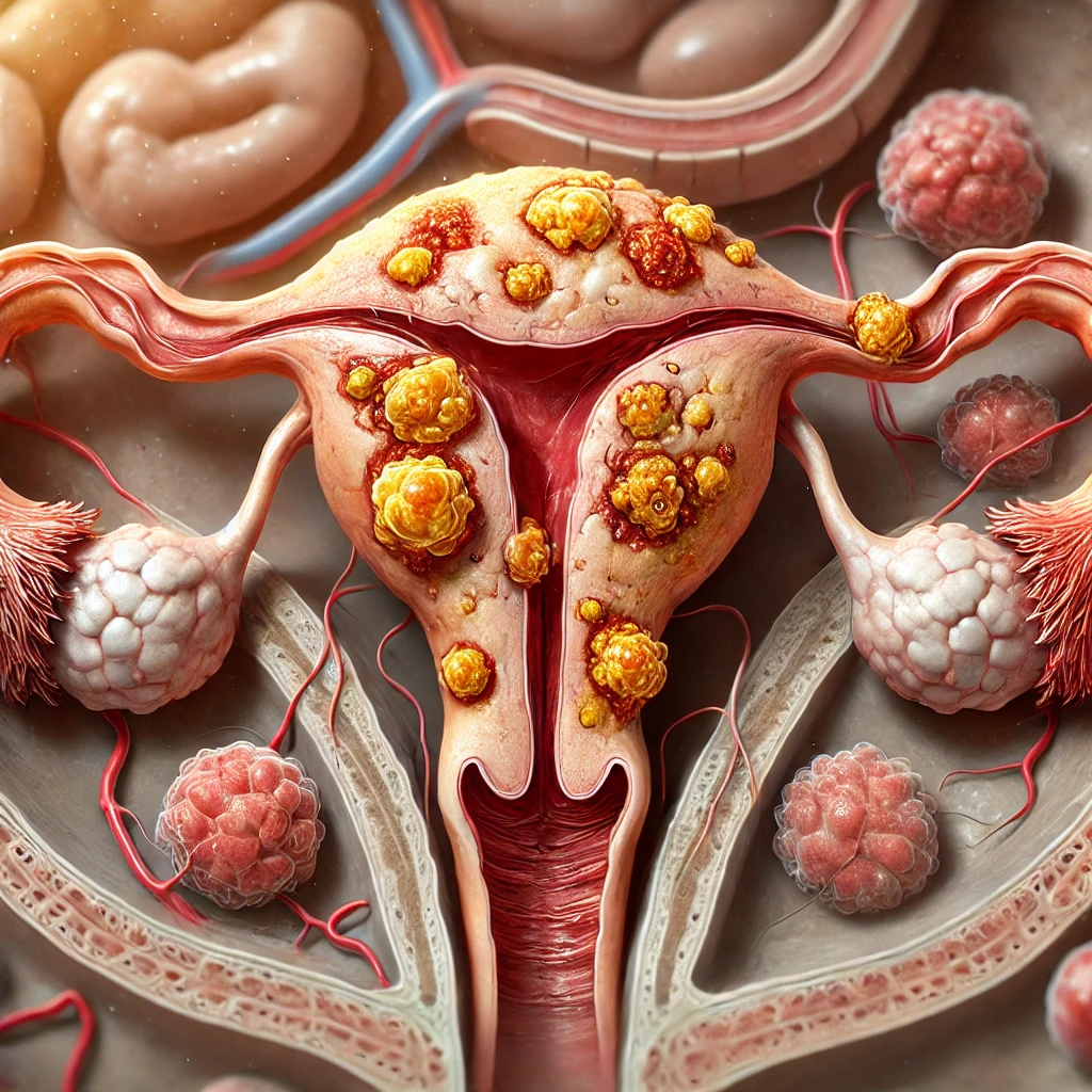
What Are Ovarian Epithelial Cancer Surface Tumors?

Ovarian epithelial cancer surface epithelial-stromal tumors develop from the cells covering the outer layer of the ovaries. These tumors represent the most common form of ovarian cancer, accounting for approximately 85 to 90 percent of malignant ovarian cases. Their classification includes three distinct types: benign, borderline, and malignant. Each type exhibits unique characteristics, influencing the tumor's behavior and potential impact on health. Understanding these tumors is essential for accurate diagnosis and effective treatment strategies.
Key Takeaways
Ovarian epithelial cancer is the most common ovarian cancer type.
It accounts for about 85-90% of all ovarian cancer cases.
These tumors can be harmless, in-between, or cancerous, needing different care.
Finding it early is important; symptoms like bloating or pain may delay diagnosis.
Knowing tumor types helps doctors create better treatment plans and save lives.
Talking to doctors about symptoms and risks can improve health results.
Overview of Ovarian Epithelial Cancer Surface Epithelial-Stromal Tumors

What Are Surface Epithelial-Stromal Tumors?
Surface epithelial-stromal tumors are a group of growths that develop from the outermost layer of the ovary, known as the ovarian surface epithelium. These tumors represent the most common type of ovarian cancer, accounting for the majority of cases. They can range from benign (non-cancerous) to borderline (low malignant potential) and malignant (cancerous). Each type exhibits distinct characteristics, influencing its behavior and treatment approach.
How Do They Originate?
These tumors originate when the cells of the ovarian surface epithelium undergo abnormal changes. Factors such as genetic mutations, hormonal influences, and environmental triggers can contribute to these changes. Individuals with a family history of ovarian, breast, or colorectal cancer face a higher risk. Inherited mutations in genes like BRCA1 and BRCA2 or conditions such as Lynch syndrome also increase susceptibility. Other risk factors include obesity, hormone therapy during menopause, and delayed or absent full-term pregnancies. Age plays a significant role, as ovarian cancer is rare in individuals under 40 but becomes more common after age 63.
Their Role in Ovarian Cancer
Ovarian epithelial cancer surface epithelial-stromal tumors play a critical role in ovarian cancer due to their prevalence and potential severity. Malignant forms of these tumors often present with vague symptoms, making early detection challenging. Common symptoms include bloating, pelvic pain, feeling full quickly, and urinary issues. These symptoms often overlap with less serious conditions, leading to delayed diagnosis. Understanding the origin and behavior of these tumors is essential for improving diagnostic accuracy and developing effective treatment strategies.
Classification of Ovarian Epithelial Cancer Surface Epithelial-Stromal Tumors

Serous Tumors
Serous tumors are the most common type of ovarian epithelial cancer surface epithelial-stromal tumor. They are categorized into benign, borderline, and malignant forms, each with distinct characteristics and implications for treatment.
Benign Serous Tumors
Benign serous tumors, also known as serous cystadenomas, are non-cancerous growths. These tumors typically present as fluid-filled cysts and rarely cause significant health issues. Surgical removal often resolves the condition, with minimal risk of recurrence.
Borderline Serous Tumors
Borderline serous tumors exhibit low malignant potential. They may spread to nearby tissues but lack invasive behavior. Approximately 60% of cases are diagnosed at Stage I, with a 5-year survival rate nearing 100%. Comprehensive surgical staging is crucial for accurate prognosis. Noninvasive implants generally do not require further therapy, while invasive implants may necessitate additional treatment discussions.
Malignant Serous Tumors
Malignant serous tumors, or serous cystadenocarcinomas, are aggressive and often diagnosed at advanced stages. These tumors frequently exhibit peritoneal implants and bilaterality, as shown in the table below:
Characteristic | Serous Tumors | Mucinous Tumors | p-value |
|---|---|---|---|
Presence of peritoneal implants | 3.6% | 0.001 | |
Positive peritoneal cytology | 35.7% | 8.5% | 0.001 |
Bilaterality | 27.9% | 1.1% | <0.0001 |
10-year Overall Survival (OS) | 98.2% | 88.5% | 0.01 |
Mucinous Tumors
Mucinous tumors are less common than serous tumors and are characterized by mucus-producing cells. They are also divided into benign, borderline, and malignant categories.
Benign Mucinous Tumors
Benign mucinous tumors, or mucinous cystadenomas, are large, fluid-filled cysts. These tumors rarely become cancerous and are often treated successfully with surgery.
Borderline Mucinous Tumors
Borderline mucinous tumors have a low risk of malignancy. They are less likely to spread compared to their serous counterparts. Proper surgical staging is essential for determining prognosis and treatment.
Malignant Mucinous Tumors
Malignant mucinous tumors, or mucinous cystadenocarcinomas, are rare but can grow significantly in size. They exhibit lower rates of bilaterality and peritoneal implants compared to malignant serous tumors. Early detection improves survival outcomes.
Endometrioid Tumors
Endometrioid tumors account for a smaller percentage of ovarian epithelial cancer surface epithelial-stromal tumors. These tumors often arise in association with endometriosis or genetic mutations. Lynch syndrome genes and BRCA1/BRCA2 mutations are implicated in their development, as shown below:
Mutation Type | Number of Cases | Percentage |
|---|---|---|
Lynch syndrome genes | 39 | 2.4% |
BRCA1 or BRCA2 | 20 | 1.2% |
Endometrioid tumors are typically malignant and require prompt intervention. Early-stage detection significantly improves prognosis.
Clear Cell Tumors
Clear cell tumors represent a rare subtype of ovarian epithelial cancer surface epithelial-stromal tumors. They account for approximately 6% of epithelial ovarian cancers in the United States. These tumors are often associated with endometriosis and typically exhibit aggressive behavior. Clear cell carcinomas are more likely to be diagnosed at an advanced stage, which complicates treatment and impacts survival rates.
Patients with clear cell tumors may experience symptoms such as abdominal pain, bloating, or pelvic discomfort. These symptoms often overlap with other ovarian tumor types, making early detection challenging. Clear cell tumors are characterized by their unique histological features, including clear cytoplasm and hobnail cells. These features help pathologists differentiate them from other ovarian tumor subtypes.
Treatment for clear cell tumors often involves a combination of surgery and chemotherapy. However, these tumors tend to exhibit resistance to standard platinum-based chemotherapy, necessitating alternative therapeutic approaches. Ongoing research aims to identify targeted therapies to improve outcomes for patients with this aggressive tumor type.
Brenner Tumors
Brenner tumors are a rare form of ovarian epithelial cancer surface epithelial-stromal tumors. They are typically benign but can also present as borderline or malignant. Borderline Brenner tumors exhibit unique histological features, including papillary structures with a fibro-vascular core covered by transitional epithelium. These tumors show specific immunohistochemical markers, such as strong EGFR expression and diffuse staining for CK7, CA125, thrombomodulin, and EMA.
Marker | Borderline Brenner Tumor | Malignant Brenner Tumor |
|---|---|---|
p16 | Negative | Negative |
Rb | Negative | Negative |
p53 | Negative | Negative |
Cyclin D1 | Weak | Strong |
Ras | Moderate | Strong |
EGFR | Strong | Strong |
p63 | Positive | Negative |
CK7 | Diffuse | Diffuse |
CA125 | Diffuse | Diffuse |
Thrombomodulin | Diffuse | Diffuse |
EMA | Diffuse | Diffuse |
Malignant Brenner tumors are extremely rare and often require aggressive treatment. Early detection and accurate diagnosis are crucial for effective management.
Undifferentiated Tumors
Undifferentiated tumors are among the most challenging ovarian epithelial cancer surface epithelial-stromal tumors to diagnose. These tumors lack distinct cellular characteristics, making it difficult to determine their lineage. They often appear primitive and less specialized, which complicates their identification. This issue is particularly pronounced in metastatic cases, where the tumors lose their original morphological and molecular markers.
Diagnosing undifferentiated tumors requires meticulous sampling and advanced diagnostic techniques. Pathologists rely on immunohistochemical and molecular markers to identify any remaining features that may indicate the tumor's origin. However, the absence of terminal differentiation often limits the effectiveness of these methods. Patients with undifferentiated tumors typically face a poor prognosis due to the aggressive nature of these cancers and the challenges in developing targeted treatment strategies.
Importance of Classification in Ovarian Epithelial Cancer
Role in Accurate Diagnosis
Classification plays a pivotal role in improving the accuracy of ovarian cancer diagnoses. Advanced diagnostic tools, such as the CNN-CAE model, have revolutionized the ability to differentiate between various types of ovarian tumors. This model achieves an impressive 97% accuracy rate, significantly outperforming traditional diagnostic methods. By eliminating visual disturbances, such as calipers and annotations in ultrasound images, it ensures a more precise evaluation of ovarian tumors. Even less experienced examiners can achieve high diagnostic accuracy using this technology. Additionally, tools like ultrasound, CT scans, and MRIs complement classification efforts by providing detailed imaging to identify tumor characteristics. The table below highlights some of the most effective diagnostic tools:
Diagnostic Tool | Description |
|---|---|
Ultrasound | Often the first test to identify ovarian tumors, distinguishing between solid masses and fluid-filled cysts. |
CT Scan | Provides detailed cross-sectional images to determine if cancer has spread to other organs. |
MRI | Uses strong magnets to create images of the inside of the body, similar to CT scans. |
PET Scan | Utilizes radioactive glucose to identify cancerous cells based on their sugar absorption rates. |
Laparoscopy | A minimally invasive procedure allowing direct visualization of the ovaries and surrounding tissues. |
Impact on Prognosis and Survival Rates
The classification of ovarian epithelial cancer surface epithelial-stromal tumors directly impacts prognosis and survival rates. Tumors diagnosed at earlier stages often have better outcomes. For instance:
The overall five-year relative survival rate for ovarian cancers is 49.7%.
Localized cancers have a survival rate of 93%, while cancers that spread to nearby regions show a 75% survival rate.
If the cancer spreads to distant areas, the survival rate drops to 31%.
Stage 1 ovarian cancers, which account for about 20% of diagnoses, have a five-year survival rate of approximately 94%. These statistics underscore the importance of early and accurate classification in improving patient outcomes.
Guiding Personalized Treatment Plans
Accurate classification also guides the development of personalized treatment plans. Each subtype of ovarian epithelial cancer surface epithelial-stromal tumor exhibits unique behavior, requiring tailored therapeutic approaches. For example, clear cell tumors often resist standard platinum-based chemotherapy, necessitating alternative treatments. Similarly, borderline tumors may not require aggressive interventions, while malignant tumors demand comprehensive strategies involving surgery and chemotherapy. By understanding the tumor's classification, oncologists can design treatments that maximize efficacy while minimizing unnecessary side effects.
Ovarian epithelial cancer surface epithelial-stromal tumors represent a diverse group of growths originating from the ovarian surface epithelium. Their classification into benign, borderline, and malignant types highlights the complexity of these tumors and their varying impacts on health. Understanding these distinctions is vital for accurate diagnosis and effective treatment planning.
Early detection and proper classification improve survival rates and guide personalized care strategies. Individuals should consult healthcare professionals to address concerns and receive tailored medical advice. This proactive approach ensures better outcomes and enhances overall well-being.
FAQ
What are the early symptoms of ovarian epithelial cancer?
Early symptoms include bloating, pelvic pain, frequent urination, and feeling full quickly. These symptoms often mimic less serious conditions, making early detection challenging. Individuals experiencing persistent symptoms should consult a healthcare provider for evaluation.
How is ovarian epithelial cancer diagnosed?
Doctors use imaging tests like ultrasounds, CT scans, or MRIs to identify tumors. Blood tests, such as CA-125, may detect cancer markers. A biopsy confirms the diagnosis by analyzing tissue samples under a microscope.
Can ovarian epithelial cancer be prevented?
While prevention is not guaranteed, reducing risk factors helps. Maintaining a healthy weight, using oral contraceptives, and undergoing genetic counseling for BRCA mutations can lower risk. Regular check-ups improve early detection.
What treatment options are available for ovarian epithelial cancer?
Treatment depends on the tumor type and stage. Common options include surgery to remove the tumor, chemotherapy to target cancer cells, and targeted therapies for specific genetic mutations. Oncologists tailor treatment plans to individual needs.
Is ovarian epithelial cancer hereditary?
Yes, some cases are hereditary. Mutations in BRCA1, BRCA2, or Lynch syndrome genes increase risk. Individuals with a family history of ovarian, breast, or colorectal cancer should consider genetic testing and counseling for personalized risk assessment.
Note: Always consult a healthcare professional for accurate diagnosis and treatment recommendations.
See Also
Exploring The Types And Nature Of Trophoblastic Tumors
Understanding Extragonadal Germ Cell Tumors And Their Formation
A Comprehensive Overview Of Gastrointestinal Carcinoid Tumors

