What Is Pilocytic Astrocytoma and Its Symptoms

Pilocytic astrocytoma is a type of brain tumor that primarily affects children and young adults. You might find it reassuring to know that this tumor grows slowly and is usually benign, meaning it rarely spreads to other parts of the body. In fact, pilocytic astrocytomas account for about 15.6% of primary brain tumors in children and adolescents. Most cases occur before the age of 10, with diagnoses often happening between the ages of 6 and 9. While it can develop in various parts of the brain, its manageable nature makes it one of the more treatable brain tumors.
Key Takeaways
Pilocytic astrocytoma is a slow-growing brain tumor. It mostly affects kids and young adults. It is usually not cancerous and can be treated.
Common signs are long-lasting headaches, feeling sick, and vision issues. Symptoms depend on where the tumor is in the brain.
Surgery is the main treatment and removes the tumor. This helps people live longer and lowers the chance of it coming back.
Regular check-ups and scans are important to track healing. They also help find any new tumor growth early.
Finding and treating it early can improve results a lot. It’s important to see a doctor if symptoms don’t go away.
Understanding Pilocytic Astrocytoma

Definition and Characteristics
What makes it "pilocytic"
The term "pilocytic" refers to the unique appearance of the tumor cells under a microscope. These cells often have long, hair-like fibers, which is where the name comes from. This feature helps doctors distinguish pilocytic astrocytomas from other types of brain tumors. The tumor's structure also contributes to its slow growth, making it less aggressive compared to other astrocytic tumors.
How it differs from other brain tumors
Pilocytic astrocytomas stand out because of their slow growth and benign nature. Unlike more aggressive brain tumors, these typically have well-defined boundaries. This characteristic makes them easier to remove surgically, often leading to successful treatment outcomes. While other astrocytic tumors may invade surrounding brain tissue, pilocytic astrocytomas tend to remain localized, which reduces the risk of spreading.
Common Locations
Cerebellum
The cerebellum is the most common site for pilocytic astrocytomas. This part of the brain controls balance and coordination, so tumors here often cause symptoms like clumsiness or difficulty walking.
Optic pathway
Pilocytic astrocytomas frequently develop along the optic pathway, which includes the optic nerve and hypothalamus. Tumors in this area can lead to vision problems, such as blurred or double vision, and may even cause hormonal imbalances if the hypothalamus is affected.
Brainstem and spinal cord
Although less common, these tumors can also appear in the brainstem or spinal cord. The brainstem controls essential functions like breathing and swallowing, so tumors here may cause difficulty with these activities. In the spinal cord, they can lead to pain or weakness in the arms or legs.
Pilocytic astrocytomas can develop in various parts of the central nervous system, but they are most often found in the cerebellum, optic pathway, hypothalamus, or brainstem. Studies show that 40% of these tumors occur in the cerebellum, while 23% affect the optic tract and hypothalamus.
Symptoms of Pilocytic Astrocytoma

General Symptoms
Pilocytic astrocytoma often presents with symptoms that can vary depending on the tumor's size and location. However, some general symptoms are commonly reported:
Persistent headaches that may worsen in the morning or with physical activity.
Nausea and vomiting, often linked to increased pressure inside the skull.
Fatigue and weakness, which can make daily activities feel more challenging.
Other symptoms you might notice include changes in personality, drowsiness, or even seizures. These signs occur because the tumor can disrupt normal brain function.
Symptoms by Tumor Location
The symptoms of pilocytic astrocytoma can also depend on where the tumor develops in the brain or spinal cord. Here's a breakdown of common symptoms based on tumor location:
Tumor Location | Symptoms |
|---|---|
Cerebellum | Difficulty with balance and coordination (ataxia), tremors, involuntary eye movements (nystagmus). |
Optic Pathway | Vision problems like blurred or double vision, loss of vision, and hormonal imbalances. |
Brainstem | Trouble swallowing or speaking, weakness on one side of the body (hemiparesis), and sensory deficits. |
Spinal Cord | Pain or weakness in the limbs, back pain, and changes in bowel or bladder function. |
For example, if the tumor is in the cerebellum, you might experience clumsiness or difficulty walking. Tumors along the optic pathway can lead to vision issues, while those in the brainstem may affect vital functions like swallowing. Spinal cord tumors often cause pain or weakness in the arms or legs.
Note: Symptoms like headaches, nausea, and vomiting are often early warning signs. If you notice these symptoms persisting, consult a healthcare professional promptly.
Causes and Risk Factors
Genetic Factors
Role of NF1 (Neurofibromatosis Type 1)
Genetic factors play a significant role in the development of pilocytic astrocytoma, especially in individuals with neurofibromatosis type 1 (NF1). If you or someone you know has NF1, the risk of developing this tumor increases. This condition stems from mutations in the NF1 gene, which produces a protein called neurofibromin. Neurofibromin acts as a growth regulator for astrocytes, the cells that form these tumors. When the NF1 gene is not functioning properly, it can lead to uncontrolled cell growth, increasing the likelihood of pilocytic astrocytoma.
People with NF1 have a higher incidence of juvenile pilocytic astrocytomas.
Loss of NF1 gene expression is directly linked to tumor development.
Neurofibromin, the NF1 gene product, helps suppress abnormal growth in astrocytes.
If you have a family history of NF1, it’s essential to monitor for symptoms and consult a healthcare provider for regular check-ups.
Sporadic Cases
Cases with no clear cause
Most cases of pilocytic astrocytoma occur sporadically, meaning they arise without a known genetic or environmental trigger. These cases are the most common type of brain tumor in children, accounting for about 25% of primary brain tumors. They typically appear between the ages of 5 and 10 and often have a favorable prognosis. However, recurrences can happen, so long-term follow-up is crucial.
Sporadic cases often involve specific genetic alterations that drive tumor growth:
The BRAF-KIAA1549 fusion is the most frequent genetic change.
Some tumors show the BRAF V600E mutation, which promotes abnormal cell growth.
Alterations in the FGFR1 gene and chromosomal abnormalities are also observed in certain cases.
These tumors commonly develop in the cerebellum, optic tract, hypothalamus, or cerebral hemispheres. While the exact cause remains unknown, understanding these genetic changes helps doctors develop targeted treatments.
Tip: Even if no clear cause is identified, early detection and treatment significantly improve outcomes for pilocytic astrocytoma.
Diagnosing Pilocytic Astrocytoma
Medical History and Physical Examination
Diagnosing pilocytic astrocytoma begins with a thorough review of your medical history and a physical examination. Your doctor will ask about symptoms like headaches, nausea, or vision problems. They may also check for neurological signs, such as difficulty with balance or coordination. These initial steps help identify potential issues and guide further testing.
Imaging Tests
MRI as the primary diagnostic tool
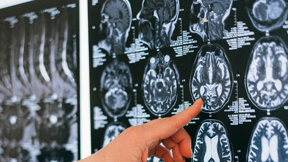
Magnetic resonance imaging (MRI) is the most effective tool for diagnosing pilocytic astrocytoma. It provides detailed images of the brain, allowing doctors to assess the tumor's size, location, and impact on surrounding tissues. Pilocytic astrocytomas often appear as well-defined, cystic masses with a solid nodule that enhances when gadolinium contrast is used. MRI can also detect complications like hydrocephalus or swelling in nearby brain tissue. Advanced techniques, such as MR spectroscopy and diffusion-weighted imaging, offer additional insights into the tumor's metabolic activity and cell density.
CT scans for additional insights
Computed tomography (CT) scans are used when MRI is not an option, such as in cases involving metal implants or severe claustrophobia. While CT scans provide less detailed images, they can identify calcifications within the tumor, although these are rare in pilocytic astrocytomas. CT scans also help evaluate the tumor's proximity to critical structures in the brain.
Biopsy for Confirmation
When and why a biopsy is performed
A biopsy is performed when imaging alone cannot confirm the diagnosis or when the tumor's location makes complete removal risky. During this minimally invasive procedure, a needle is guided into the tumor to collect a small tissue sample. Pathologists examine the sample under a microscope, looking for specific features like Rosenthal fibers and dense cell areas. Molecular tests may also detect genetic changes, such as the BRAF-KIAA1549 fusion, which is common in pilocytic astrocytomas.
While biopsies are generally safe, they carry some risks, including neurological deficits or functional impairments, especially if the tumor is in sensitive areas like the brainstem. However, the information gained from a biopsy is crucial for confirming the diagnosis and planning effective treatment.
Treatment and Prognosis
Surgery
Complete resection as the goal
Surgery is often the first-line treatment for pilocytic astrocytoma. The primary goal is complete resection, where the entire tumor is removed. This approach significantly improves long-term outcomes.
Gross total resection (GTR) is associated with excellent survival rates.
The 10-year postoperative survival rate can reach up to 95.8%.
Complete removal reduces the risk of recurrence, offering a favorable prognosis.
Risks and recovery
While surgery is highly effective, it carries potential risks, especially when the tumor is located in sensitive areas like the brainstem or ventricles. Complications may include functional impairments, memory deficits, or language difficulties. Extensive resection in high-risk areas can also lead to significant blood loss, impacting recovery. After surgery, you may need regular follow-ups to monitor for recurrence and manage any lasting effects. Emotional support and rehabilitation can also play a vital role in recovery.
Radiation Therapy
When surgery is not fully effective
Radiation therapy becomes an option when the tumor cannot be completely removed or if it recurs after surgery. It targets remaining tumor cells to prevent further growth. However, radiation therapy carries long-term risks, particularly for children. Studies show it may lead to cognitive deficits, hormonal imbalances, and even an increased risk of malignant transformation.
Note: Doctors carefully weigh the benefits and risks of radiation therapy, especially for younger patients.
Description | |
|---|---|
Cognitive deficits | Impairments in memory, attention, and learning abilities. |
Endocrine dysfunctions | Hormonal imbalances affecting growth and metabolism. |
Malignant transformation risk | Increased likelihood of tumor becoming cancerous due to radiation exposure. |
Chemotherapy
Use in specific cases, such as inoperable tumors
Chemotherapy is typically reserved for cases where surgery and radiation are not viable options. It is often used for inoperable tumors or when the tumor progresses despite other treatments. Studies show that chemotherapy can stabilize or improve symptoms in some patients.
Number of Patients | Percentage | |
|---|---|---|
Clinical and radiographic improvement (R/R) | 4 | 36% |
Clinical stabilization and radiographic improvement (SD/R) | 1 | 9% |
Clinical and radiographic stabilization (SD/SD) | 3 | 27% |
Clinical and radiographic progression (PD/PD) | 3 | 27% |
Doctors may use various chemotherapy regimens, tailoring the treatment to your specific needs. While chemotherapy can be effective, it often requires close monitoring to manage side effects and ensure the best possible outcome.
Prognosis and Long-Term Outcomes
High survival rates with proper treatment
Pilocytic astrocytoma has one of the most favorable prognoses among pediatric brain tumors. With timely diagnosis and effective treatment, survival rates are exceptionally high. For instance, the 5-year survival rate reaches 100%, while the 10-year survival rate stands at 95.8%.
Time After Diagnosis | Survival Rate |
|---|---|
5 years | 100% |
10 years | 95.8% |
Complete surgical resection plays a critical role in achieving these outcomes. When surgeons successfully remove the entire tumor, the chances of recurrence drop significantly. Younger patients often experience better results due to their ability to recover more effectively from surgery. Compared to other pediatric brain tumors, pilocytic astrocytoma offers a much brighter outlook, especially when treated early.
Tip: Regular follow-ups after treatment ensure any signs of recurrence are detected early, further improving long-term outcomes.
Potential for recurrence and follow-up care
Although pilocytic astrocytoma is typically benign, recurrence remains a possibility, especially if the tumor cannot be fully removed. Several factors influence the likelihood of recurrence:
The extent of surgical resection is the most important factor. Gross total resection (GTR) significantly reduces recurrence risk.
Tumor location matters. Tumors in accessible areas, like the cerebellum, are easier to remove completely, while those in the optic pathway or brainstem pose challenges.
Younger patients often have better outcomes due to improved surgical success rates.
Conditions like neurofibromatosis type 1 (NF1) can complicate the disease and increase recurrence risk.
Follow-up care is essential for managing these risks. Regular imaging tests, such as MRIs, help monitor for any signs of tumor regrowth. If recurrence occurs, additional treatments like radiation therapy or chemotherapy may be necessary. Maintaining overall health and attending routine check-ups can also improve long-term outcomes.
Note: Early detection of recurrence allows for prompt intervention, which can prevent complications and improve quality of life.
Pilocytic astrocytoma is a slow-growing brain tumor that often affects children and young adults. You’ve learned that its symptoms depend on the tumor’s location, with common signs including headaches, nausea, and vision problems. Treatment options like surgery, radiation therapy, and chemotherapy offer effective ways to manage this condition.
The prognosis for pilocytic astrocytoma is highly encouraging. Over 90% of children diagnosed with this tumor survive beyond five years, and complete tumor removal significantly lowers recurrence rates. Early detection through imaging tests and medical evaluations plays a vital role in achieving these outcomes.
If you notice persistent symptoms, consult a healthcare professional promptly. Early diagnosis and treatment can make a significant difference in ensuring a healthy future.
FAQ
What is the difference between pilocytic astrocytoma and other brain tumors?
Pilocytic astrocytoma grows slowly and usually remains localized. Unlike aggressive brain tumors, it has well-defined boundaries, making surgical removal easier. Its benign nature means it rarely spreads to other parts of the body.
Can pilocytic astrocytoma recur after treatment?
Yes, recurrence is possible, especially if the tumor isn’t fully removed. Regular follow-ups and imaging tests help detect regrowth early. Complete surgical removal significantly lowers the risk of recurrence.
Are there any long-term effects of pilocytic astrocytoma treatment?
Some treatments, like radiation therapy, may cause cognitive or hormonal changes, especially in children. Surgery can also lead to temporary neurological issues. Regular monitoring and rehabilitation minimize these effects.
How is pilocytic astrocytoma diagnosed?
Doctors use MRI scans to identify the tumor’s size and location. A biopsy confirms the diagnosis by analyzing the tumor’s cellular structure. These steps ensure accurate identification and treatment planning.
Is pilocytic astrocytoma life-threatening?
Pilocytic astrocytoma is rarely life-threatening with timely diagnosis and treatment. Most patients, especially children, have excellent survival rates. Early intervention and complete tumor removal improve outcomes significantly.
Tip: Always consult a healthcare professional if you notice persistent symptoms like headaches or vision problems. Early detection makes a big difference.
See Also
Understanding Cerebellar Astrocytoma: Key Symptoms To Recognize
Ependymoma Explained: Recognizing Its Symptoms and Effects
Chondrosarcoma Overview: Identifying Symptoms You Should Know
Cerebral Astrocytoma: Impact on Brain Function and Symptoms
Choroid Plexus Carcinoma: Symptoms and Treatment Options Explained

