What is Glioma and Its Types

Glioma is a type of tumor that starts in the brain or spinal cord. It develops from glial cells, which support and protect nerve cells in your central nervous system. This tumor is the most common type found in the brain and spinal cord. Its prevalence makes it a critical topic for understanding brain health. Recognizing its impact can help you stay informed about potential risks.
Key Takeaways
Gliomas are growths that start in brain or spine cells. Knowing their types and levels helps understand their effects on health.
Finding gliomas early can make treatments work better. Watch for signs like constant headaches or seizures and see a doctor quickly.
Types of gliomas, like astrocytomas and glioblastomas, are different. Learning about them helps in making good health choices.
Regular doctor visits and scans help find gliomas early. Taking care of brain health can lower possible risks.
New treatments, like immunotherapy and targeted drugs, give hope. Staying updated on new ideas can lead to better care.
What is Glioma?
Definition and Characteristics
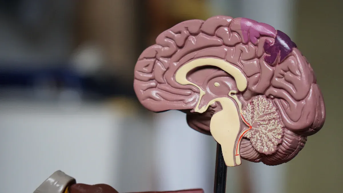
Glioma refers to a tumor that arises from glial cells in your brain or spinal cord. These cells play a crucial role in supporting and protecting neurons. Gliomas differ from other brain tumors due to their unique characteristics, which vary based on their grade. The table below outlines these differences:
Grade | Characteristics |
|---|---|
1 | Slow-growing, almost normal-looking cells, well-defined boundaries, noncancerous but can cause symptoms. |
2 | Slow-growing, more abnormal cells, infiltrates nearby tissue, potential to become cancerous. |
3 | Rapid growth, invasive, malignant, higher recurrence rate. |
4 | Most aggressive, fast-growing, abnormal cells, often spreads within the brain, glioblastoma is the most common form. |
Understanding these grades helps you recognize how gliomas progress and why early detection is essential.
How Gliomas Form
Gliomas form when glial cells undergo abnormal changes. These changes disrupt their normal function and cause uncontrolled cell growth. Factors like genetic mutations and environmental influences can trigger this process. As the tumor grows, it may invade nearby tissues, leading to various symptoms. The exact cause of gliomas remains unclear, but researchers continue to study their development to improve treatment options.
Why Gliomas Are Common in the Central Nervous System
Gliomas are the most common tumors in the central nervous system because glial cells are abundant in your brain and spinal cord. These cells outnumber neurons and perform essential tasks like providing nutrients, maintaining the blood-brain barrier, and repairing damage. Their high numbers and active role in cell division make them more susceptible to mutations, increasing the likelihood of tumor formation.
Types of Gliomas

Astrocytomas
Astrocytomas develop from astrocytes, which are star-shaped cells in your central nervous system. These tumors can occur in both the brain and spinal cord. Symptoms often include headaches, blurry vision, seizures, and mood changes. The treatment approach depends on the tumor's grade:
Grade 1 astrocytomas grow slowly and may not require further treatment after surgery.
Grades 3 and 4 are more aggressive and need a combination of surgery, radiation, and chemotherapy.
Higher-grade astrocytomas pose a greater challenge because they infiltrate healthy brain tissue, making complete removal difficult. Early detection and treatment can improve outcomes significantly.
Tip: If you experience persistent headaches or seizures, consult a healthcare professional promptly.
Oligodendrogliomas
Oligodendrogliomas arise from oligodendrocytes, which produce the protective myelin sheath around nerve fibers. These tumors are less common but have distinct genetic features that influence their behavior and treatment. The table below highlights key genetic mutations associated with oligodendrogliomas:
Genetic Mutation | Description |
|---|---|
Isocitrate dehydrogenase (IDH) mutation | Common in oligodendrogliomas, linked to less aggressive tumors and longer survival rates. |
Codeletion of chromosomal arms 1p and 19q | Found in all oligodendrogliomas, associated with better prognosis compared to other gliomas. |
These genetic markers help doctors tailor treatment plans and predict outcomes. Oligodendrogliomas often respond well to therapy, especially when detected early.
Ependymomas
Ependymomas can form anywhere in your central nervous system, but they often develop near the brain's ventricles or the spinal cord's central canal. These tumors vary in grade, as shown below:
Grade | Description | Characteristics |
|---|---|---|
1 | Low-grade | Subependymomas, more common in adults than children. |
2 | Low-grade | Includes myxopapillary and conventional subtypes, more likely to recur. |
3 | Malignant | Fast-growing, occurs most often in the brain. |
Ependymomas differ from other gliomas because they can occasionally form outside the central nervous system, such as in the ovaries. Symptoms depend on the tumor's location and size, but they often include nausea, balance issues, and weakness.
Note: Regular check-ups and imaging tests can help detect these tumors early, improving treatment success.
Glioblastoma (Grade IV Glioma)
Glioblastoma, also known as Grade IV glioma, is the most aggressive type of brain tumor. It falls under the category of astrocytomas but stands out due to its rapid growth and invasive nature. This tumor starts in the brain and rarely spreads outside the central nervous system. Its aggressive behavior makes it one of the most challenging gliomas to treat.
Several factors contribute to glioblastoma's severity:
The tumor consists of abnormal cells that grow and divide rapidly.
It invades nearby brain tissue, making complete surgical removal difficult.
Glioblastoma often forms new blood vessels to support its growth, further complicating treatment.
Symptoms of glioblastoma vary depending on its location in the brain. You might experience persistent headaches, nausea, or seizures. Some individuals notice changes in memory, mood, or speech. These symptoms often worsen quickly due to the tumor's fast progression.
Treatment for glioblastoma typically involves a combination of surgery, radiation therapy, and chemotherapy. Surgeons aim to remove as much of the tumor as possible without damaging healthy brain tissue. Radiation and chemotherapy target remaining cancer cells to slow the tumor's growth. Despite these efforts, glioblastoma often recurs, requiring additional treatment.
Researchers continue to explore new therapies to improve outcomes for glioblastoma patients. Emerging treatments, such as immunotherapy and targeted drug therapies, show promise in clinical trials. Early detection remains crucial. If you notice persistent neurological symptoms, consult a healthcare professional immediately.
Note: Glioblastoma's aggressive nature highlights the importance of ongoing research and advancements in treatment options.
Symptoms of Gliomas
General Symptoms
Gliomas can cause a wide range of symptoms, depending on their size and location. Some of the most common symptoms include:
Nausea and vomiting
Memory problems or confusion
Blurred or double vision
Fatigue and dizziness
Personality changes or mood disturbances
Weakness or numbness in your arms, legs, or face
These symptoms occur because the tumor disrupts normal brain or spinal cord function. For example, headaches may result from increased pressure inside your skull, while seizures happen due to abnormal electrical activity in the brain. If you notice any of these signs, it’s important to monitor their frequency and severity.
Symptoms Based on Tumor Location
The location of a glioma plays a significant role in the symptoms you experience. Tumors in different areas of the brain or spinal cord can affect specific functions. For instance:
Frontal lobe tumors may cause personality changes, memory issues, or difficulty concentrating.
Temporal lobe tumors often lead to seizures, speech problems, or hearing difficulties.
Parietal lobe tumors can result in numbness, weakness, or coordination problems.
Occipital lobe tumors may cause vision loss or disturbances.
Spinal cord tumors can lead to back pain, weakness, or difficulty walking.
Symptoms may develop slowly, making them easy to overlook at first. However, as the tumor grows, these signs often become more noticeable and severe.
When to Seek Medical Advice
You should seek medical advice if you experience persistent or worsening symptoms, especially if they interfere with your daily life. For example, recurring headaches that don’t improve with medication or new-onset seizures require immediate attention. Sudden changes in vision, speech, or coordination are also red flags. Early diagnosis can improve treatment outcomes and help manage symptoms effectively.
Tip: Keep a symptom diary to track changes over time. This information can help your doctor make an accurate diagnosis.
Diagnosis of Gliomas
Imaging Techniques
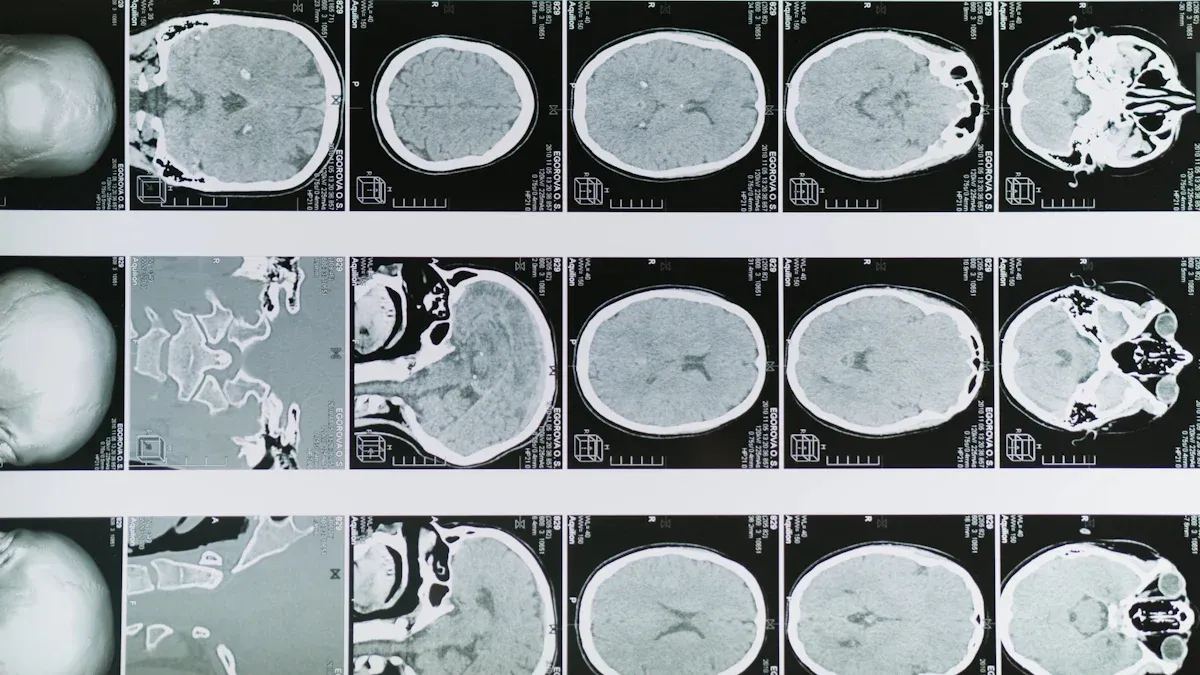
MRI
Magnetic resonance imaging (MRI) is often the first choice for diagnosing gliomas. This technique uses powerful magnets and radio waves to create detailed images of your brain or spinal cord. MRI scans can identify the tumor's size, shape, and location with high precision. Doctors may also use advanced MRI techniques, such as functional MRI, to assess how the tumor affects nearby brain functions.
CT Scans
Computed tomography (CT) scans use X-rays to produce cross-sectional images of your brain. These scans help evaluate the tumor's size and location. CT scans are particularly useful in emergency situations or when MRI is unavailable. While they provide less detail than MRI, they remain an important tool in glioma diagnosis.
Imaging Technique | Description |
|---|---|
MRI | Often the first choice in neuroimaging for gliomas. |
CT Scan | Helps evaluate tumor size and location using X-rays for detailed images. |
PET Scan | Evaluates metabolic activity of tumors to determine aggressiveness. |
Tip: MRI scans are more detailed, but CT scans can be quicker and more accessible in certain cases.
Biopsy and Histological Analysis
A biopsy is essential when imaging suggests a glioma. During this procedure, a surgeon removes a small tissue sample from the tumor. This can be done through open surgery or a minimally invasive stereotactic biopsy. Neuropathologists then examine the tissue under a microscope to determine the tumor's type and grade. This analysis helps guide your treatment plan.
Note: A biopsy provides critical information about the tumor's characteristics, which imaging alone cannot reveal.
Other Diagnostic Methods
In addition to imaging and biopsy, other diagnostic methods can provide valuable insights:
Molecular testing identifies genetic mutations that influence prognosis and treatment.
Magnetic resonance spectroscopy (MRS) analyzes the chemical composition of tumors, especially for lower-grade gliomas.
Neurological exams assess vision, hearing, balance, and reflexes to pinpoint affected brain areas.
Genetic testing detects DNA mutations driving tumor growth, aiding in personalized treatment planning.
These methods complement imaging and biopsy, offering a comprehensive understanding of your glioma.
Treatment and Prognosis of Gliomas
Treatment Options
Surgery
Surgery is often the first step in treating gliomas. It allows doctors to remove as much of the tumor as possible while preserving healthy tissue. This procedure also provides a sample for biopsy, helping to confirm the tumor type and grade. In some cases, complete removal may not be possible due to the tumor's location. However, even partial removal can reduce symptoms and improve the effectiveness of other treatments.
Radiation Therapy
Radiation therapy uses high-energy beams to destroy tumor cells. It is commonly recommended after surgery to target any remaining cancerous cells. Techniques like stereotactic radiosurgery deliver precise doses of radiation, minimizing damage to surrounding tissues. This approach can shrink tumors and reduce the risk of recurrence.
Chemotherapy
Chemotherapy involves drugs that kill cancer cells or stop their growth. Temozolomide is a common choice for gliomas, often used alongside radiation therapy. This combination improves survival rates, especially for aggressive tumors like glioblastomas. Chemotherapy can be administered orally or intravenously, depending on the treatment plan.
Emerging Therapies
Emerging therapies offer hope for more effective glioma treatments. These include:
Targeted therapy, which focuses on specific molecules involved in tumor growth.
Tumor-treating fields (TTF) therapy, using electrical fields to disrupt cancer cell division.
Immunotherapy, which boosts your immune system to fight the tumor.
While these treatments are still under research, they show promise in clinical trials.
Factors Affecting Prognosis
Several factors influence your prognosis:
Tumor grade: Higher grades are more aggressive and harder to treat.
Age: Younger patients often respond better to treatment.
Extent of tumor removal: More complete removal improves outcomes.
Overall health: A strong immune system supports recovery.
For example, Grade I gliomas have a 5-year survival rate of up to 95%, while Grade IV glioblastomas have a much lower rate, often around 5%.
Importance of Early Detection
Early detection plays a critical role in improving treatment outcomes. Identifying gliomas before they progress allows doctors to implement effective strategies, such as surgery and targeted therapies. If you notice persistent symptoms like headaches or seizures, seeking medical advice promptly can make a significant difference.
Tip: Regular check-ups and imaging tests can help detect gliomas early, especially if you have risk factors.
Glioma, a tumor originating from glial cells in the brain or spinal cord, includes various types such as astrocytomas, oligodendrogliomas, ependymomas, and glioblastomas. Recognizing its symptoms early, like persistent headaches or seizures, can lead to timely diagnosis and better outcomes. You should always prioritize your health by consulting a medical professional if you notice concerning signs.
To learn more about gliomas, explore these resources:
Informing Health: A Guide to Healthcare Information
Advancements in Brain Cancer Treatment
Ivy Brain Tumor Center - New Treatments
Brain Cancer Breakthrough: New Clinical Trial Tests Direct-to-Tumor Method
Public health strategies also play a vital role in raising awareness:
Campaigns provide accurate information to patients at diagnosis.
Patients learn about clinical trial options early.
Advanced biomarker testing promotes targeted therapies.
By staying informed and proactive, you can take control of your health and support ongoing advancements in glioma care.
FAQ
What causes gliomas to develop?
Gliomas develop when glial cells undergo abnormal changes, leading to uncontrolled growth. Genetic mutations and environmental factors, like radiation exposure, may trigger these changes. While the exact cause remains unclear, researchers believe a combination of genetic predisposition and external influences plays a role.
Are gliomas hereditary?
Most gliomas are not hereditary. However, certain genetic conditions, like Li-Fraumeni syndrome or neurofibromatosis, can increase your risk. If you have a family history of brain tumors, discussing genetic testing with your doctor may help assess your risk.
Can gliomas be cured?
Low-grade gliomas may be curable with surgery, especially if detected early. High-grade gliomas, like glioblastomas, are more challenging to treat. While a complete cure is rare, treatments like surgery, radiation, and chemotherapy can manage symptoms and improve quality of life.
How can you reduce your risk of gliomas?
You can reduce your risk by avoiding unnecessary radiation exposure and maintaining a healthy lifestyle. While no guaranteed prevention exists, staying informed about symptoms and seeking early medical advice can help detect gliomas sooner.
Tip: Regular check-ups and a balanced diet support overall brain health.
What is the survival rate for gliomas?
Survival rates depend on the glioma's type and grade. For example, Grade I gliomas have a 5-year survival rate of up to 95%. Glioblastomas (Grade IV) have a much lower rate, often around 5%. Early detection and treatment improve outcomes significantly.
Note: Discuss your specific prognosis with your healthcare provider for accurate information.
---
ℹ️ Explore more: Read our Comprehensive Guide to All Known Cancer Types for symptoms, causes, and treatments.
See Also
Exploring The Different Types Of Brainstem Glioma
A Comprehensive Guide To The Types Of Astrocytoma
An Overview Of Blastoma And Its Various Types
Essential Information About Ganglioneuroma You Must Understand

© 2026 Banish Cancer. Banish Cancer is a registered non-profit organization providing evidence-based research, educational courses, and Fear Response AI support services. Reg. No: 305706884 | Address: Taikos pr. 43-603, Kaunas, Lithuania.