What is Oligodendroglioma and How is it Treated

Oligodendroglioma is a rare brain tumor that develops in oligodendrocytes, the cells responsible for supporting nerve fibers in your central nervous system. It accounts for about 1.4% of all primary brain tumors, with an occurrence rate of 0.2 per 100,000 people. In the United States, approximately 14,950 individuals live with this condition. Early diagnosis plays a critical role in improving outcomes. Imaging tests like MRIs and biopsies help confirm the diagnosis, while identifying genetic markers such as the 1p/19q co-deletion can guide treatment strategies.
Key Takeaways
Oligodendroglioma is a rare brain tumor that starts in brain cells. Finding it early with tests like MRIs can help treatment work better.
There are two types of oligodendroglioma: Grade II tumors grow slowly and may not change much, while Grade III tumors grow quickly and need stronger treatment.
Common signs include bad headaches, seizures, and trouble thinking clearly. Spotting these symptoms early can help doctors act fast.
Treatment usually includes surgery to take out the tumor. This is often followed by radiation and chemotherapy. Each patient needs a plan that fits them best.
How well someone does depends on the tumor type and genes. Slow-growing tumors often mean longer survival, so check-ups and support are very important.
What is Oligodendroglioma?

Definition and Overview
Oligodendroglioma is a type of brain tumor that originates from oligodendrocytes, the cells responsible for insulating and supporting nerve fibers in your central nervous system. These tumors are a subtype of gliomas and account for about 10% of all glioma diagnoses. Compared to other gliomas, oligodendrogliomas grow more slowly and respond better to treatment. They often occur in the frontal lobes of the brain, where they may cause symptoms like headaches or seizures. Specific genetic markers, such as IDH mutations and the 1p/19q codeletion, help distinguish oligodendrogliomas from other types of brain tumors.
Types of Oligodendroglioma
Grade II (Low-Grade)
Grade II oligodendrogliomas grow slowly and typically have a less aggressive nature. These tumors appear slightly abnormal under a microscope and may remain stable for years. However, they can progress to a more aggressive form over time. Treatment often focuses on surgical removal, aiming to achieve maximal safe resection.
Grade III (Anaplastic)
Grade III oligodendrogliomas, also known as anaplastic oligodendrogliomas, grow more rapidly and exhibit more abnormal features. These tumors may spread within the brain or spinal cord. Treatment for Grade III tumors usually involves a combination of surgery, chemotherapy, and radiation therapy. This aggressive approach helps manage the tumor's faster progression and higher risk of recurrence.
Grade | Growth Rate | Appearance | Progression |
|---|---|---|---|
II | Slow | Slightly abnormal | Can progress to Grade III |
III | Fast | More abnormal | May present as aggressive early |
Causes and Risk Factors
Genetic Mutations (e.g., IDH mutation, 1p/19q co-deletion)
Oligodendrogliomas are closely linked to specific genetic changes. To confirm a diagnosis, doctors look for two key alterations: an IDH mutation and the 1p/19q codeletion. The IDH mutation is associated with less aggressive tumors and longer survival rates. The 1p/19q codeletion, found in all oligodendrogliomas, indicates a better prognosis compared to other gliomas.
Genetic Mutation | Description |
|---|---|
IDH mutation | Found in most oligodendrogliomas; linked to slower growth and better outcomes. |
1p/19q codeletion | Essential for diagnosis; associated with improved survival and treatment response. |
Environmental and Lifestyle Factors
While genetic mutations play a significant role, environmental and lifestyle factors may also contribute to the risk of developing oligodendroglioma. For example, exposure to ionizing radiation, such as radiation therapy during childhood, increases the likelihood of brain tumors. Although rare, a family history of gliomas or hereditary cancer syndromes like Li-Fraumeni syndrome may also raise your risk. Some inherited gene variants have been linked to a slightly higher chance of developing these tumors.
Risk Factor | Description |
|---|---|
Exposure to Ionizing Radiation | Higher risk for individuals who had radiation therapy, especially during childhood. |
Family History | Rarely runs in families; genetic components may play a role in some cases. |
Hereditary Cancer Syndromes | Conditions like Li-Fraumeni syndrome can lead to early onset of multiple tumors, including gliomas. |
Inherited Gene Variants | Certain variants slightly increase risk but do not guarantee tumor development. |
Symptoms of Oligodendroglioma
Common Symptoms
Oligodendroglioma often presents with symptoms that vary depending on the tumor's size and location. However, some symptoms are frequently reported:
Headaches: You may experience persistent headaches that worsen over time. These headaches can sometimes wake you up at night.
Seizures: Seizures are often the first noticeable sign. They may occur suddenly, even if you have no prior history of epilepsy.
Cognitive or Behavioral Changes: Memory problems, confusion, or difficulty concentrating are common. You might also notice mood disturbances or changes in personality.
Weakness or Numbness in Limbs: Tumors can affect motor control, leading to muscular weakness or numbness, often on one side of the body.
Other symptoms include blurred vision, fatigue, and balance problems. These symptoms may develop gradually, making early detection challenging.
Symptoms Based on Tumor Location
The symptoms of oligodendroglioma can also depend on where the tumor is located in your brain.
Frontal Lobe Symptoms
If the tumor is in the frontal lobe, you might notice changes in personality or behavior. This area of the brain controls decision-making and emotions, so you could feel unusually irritable or have difficulty planning tasks. Weakness in body movements, especially on one side, is another common sign.
Temporal Lobe Symptoms
A tumor in the temporal lobe can cause seizures or speech problems. You might also experience altered sensations, such as strange smells or auditory hallucinations. Memory issues and difficulty understanding language are other potential symptoms.
The location of the tumor plays a significant role in determining the symptoms you experience. Understanding these signs can help you seek medical attention promptly.
How is Oligodendroglioma Diagnosed?
Initial Evaluation
Medical History and Physical Exam
Your diagnosis begins with a detailed medical history and physical exam. Doctors ask about your symptoms, their duration, and any family history of brain tumors or genetic conditions. They also perform a physical exam to check for visible signs of neurological issues. This step helps identify potential risk factors and narrows down the possible causes of your symptoms.
Neurological Assessment
A neurological assessment evaluates how well your brain and nervous system function. During this exam, doctors test your reflexes, balance, coordination, muscle strength, vision, and hearing. These tests help pinpoint areas of the brain that may be affected by the tumor.
Imaging Tests
MRI (Magnetic Resonance Imaging)
MRI is the most effective imaging tool for diagnosing oligodendroglioma. It provides detailed images of your brain, showing the tumor's size, location, and characteristics. Advanced techniques like dynamic contrast-enhanced MRI can assess the tumor's microvasculature and help differentiate between low-grade and high-grade tumors. This information is crucial for planning treatment.
CT Scan
Doctors may use a CT scan as an initial imaging test, especially if you have nonspecific symptoms. While CT scans are less detailed than MRIs, they can quickly detect abnormalities in your brain. However, MRI remains superior for characterizing oligodendrogliomas, particularly when the tumor involves the brain's cortex.
Biopsy and Pathological Analysis
Tissue Sampling
A biopsy involves removing a small sample of the tumor tissue for microscopic examination. This step confirms the diagnosis and determines the tumor's grade. Surgeons use advanced techniques to ensure the biopsy is as safe and accurate as possible.
Genetic Testing for 1p/19q Co-deletion
Genetic testing plays a vital role in diagnosing oligodendroglioma. Doctors analyze the tumor tissue for specific genetic changes, such as the 1p/19q co-deletion and IDH mutations. These markers confirm the diagnosis and guide treatment decisions. For example, the presence of the 1p/19q co-deletion indicates a better prognosis and helps predict how well the tumor will respond to therapy.
Treatment Options for Oligodendroglioma

Surgery
Goal of Surgery (e.g., Tumor Removal or Reduction)
Surgery is often the first step in treating oligodendroglioma. The primary goal is to remove as much of the tumor as possible while preserving brain function. In some cases, surgeons aim for complete removal, which can improve outcomes, especially for low-grade tumors. If complete removal isn’t possible, partial resection helps reduce symptoms and provides tissue for diagnosis. This approach allows doctors to confirm the tumor type and plan further treatment.
Risks and Recovery
Modern surgical techniques minimize risks and improve recovery times. For example, microsurgery uses a microscope for precision, reducing trauma to surrounding brain tissue. Endoscopic neurosurgery involves small, flexible tools, making it less invasive and promoting faster recovery. The table below highlights these techniques:
Surgical Technique | Description | Recovery Implications |
|---|---|---|
Microsurgery | Uses a microscope for precision in operating on small brain structures. | Faster recovery due to less trauma. |
Endoscopic neurosurgery | Involves small, flexible tubes for visualization and access. | Minimally invasive, leading to quicker recovery. |
Recovery depends on the tumor’s location and the extent of resection. You may experience temporary fatigue or neurological symptoms, but most patients recover well with proper care.
Radiation Therapy
When Radiation is Used
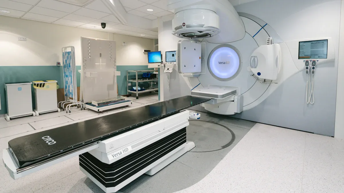
Radiation therapy is often recommended after surgery, especially for high-grade or partially removed tumors. It helps destroy remaining cancer cells and reduces the risk of recurrence. Doctors may also use radiation as a primary treatment if surgery isn’t an option.
Types of Radiation Therapy
Several advanced radiation techniques target the tumor while sparing healthy tissue. These include:
Intensity-Modulated Radiation Therapy (IMRT): Uses 3D imaging to focus radiation precisely on the tumor.
Image-Guided Radiation Therapy (IGRT): Utilizes real-time imaging for accurate radiation delivery.
Stereotactic Radiosurgery: Delivers a high dose of radiation directly to the tumor, often used for small or hard-to-reach tumors.
Type of Radiation Therapy | Description |
|---|---|
Intensity-Modulated Radiation Therapy | Uses advanced software and 3-D imaging to focus radiation on the tumor, minimizing exposure to healthy tissue. |
Image-Guided Radiation Therapy | Utilizes real-time imaging to ensure accurate delivery of radiation during treatment. |
Stereotactic Radiosurgery | Delivers a high dose of radiation precisely to the tumor, often used as an alternative to open surgery. |
Chemotherapy
Common Drugs (e.g., Temozolomide, PCV)
Chemotherapy is another key treatment for oligodendroglioma. Drugs like Temozolomide and PCV (Procarbazine, Lomustine, and Vincristine) are commonly used. These medications slow tumor growth and improve survival rates. For tumors with IDH mutations, targeted therapies like Vorasidinib have shown promise. Taken once daily, Vorasidinib slows tumor regrowth and offers a new option for treating grade II oligodendrogliomas.
Side Effects and Management
Chemotherapy can cause side effects such as nausea, fatigue, and hair loss. You can manage these symptoms with supportive care, including anti-nausea medications and dietary adjustments. Regular follow-ups with your doctor ensure that side effects are addressed promptly, helping you maintain a better quality of life during treatment.
Targeted Therapies and Emerging Treatments
Experimental Therapies
Targeted therapies focus on specific genetic mutations or pathways involved in tumor growth. For oligodendroglioma, researchers are exploring treatments that target IDH1 and IDH2 mutations. These therapies aim to slow tumor progression and improve survival rates. Vorasidinib, recently approved for grade II oligodendrogliomas with IDH mutations, has shown promise in delaying tumor regrowth. This breakthrough offers hope for patients with slow-growing tumors.
Immunotherapies are another exciting area of research. These treatments harness your immune system to fight cancer. Checkpoint inhibitors and CAR T-cell therapy are being tested for their ability to attack tumor cells while sparing healthy tissue. Although still experimental, these approaches could revolutionize how oligodendrogliomas are treated in the future.
Clinical Trials
Clinical trials provide access to cutting-edge treatments under investigation. If you have oligodendroglioma, participating in a trial might offer new options when standard treatments are insufficient. Current trials include therapies targeting IDH mutations and other innovative approaches:
These trials also explore molecular targeted therapies and immunotherapies. By joining a trial, you contribute to advancing medical knowledge while potentially benefiting from innovative treatments.
Note: Always consult your doctor before considering experimental therapies or clinical trials. They can help determine if these options are right for you.
Prognosis and Survival Rates for Oligodendroglioma
Factors Influencing Prognosis
Tumor Grade and Genetic Markers
The grade of the tumor and its genetic markers significantly impact your prognosis. Grade 2 tumors grow slowly and have a better outlook, while grade 3 (anaplastic) tumors are more aggressive and require intensive treatment. Genetic markers like the IDH mutation and 1p/19q codeletion also play a critical role. These markers predict how your tumor will behave and respond to treatment. For example:
IDH mutations indicate a less aggressive tumor and better survival rates.
The 1p/19q codeletion is associated with improved outcomes compared to other gliomas.
Patient Age and Overall Health
Your age and overall health also influence your prognosis. Patients under 60 years old tend to have better outcomes. A lower Ki-67 index, which measures tumor cell proliferation, is another positive factor. Additionally, the absence of symptoms like dementia or malignancy improves your chances of a favorable prognosis.
Survival Rates
Low-Grade Oligodendroglioma
Low-grade oligodendrogliomas generally have higher survival rates. The 1-year survival rate is 94.5%, and the 5-year survival rate is 82.7%. These tumors grow slowly, allowing for longer progression-free survival. However, regular monitoring remains essential to detect any changes.
Anaplastic Oligodendroglioma
Anaplastic oligodendrogliomas have a more aggressive nature, leading to lower survival rates. The median progression-free survival is 19 months, and the median overall survival is 37.7 months. The table below highlights survival rates for both types:
Type of Oligodendroglioma | 1-Year Survival Rate | 2-Year Survival Rate | 5-Year Survival Rate |
|---|---|---|---|
Anaplastic | 85.8% | 74.3% | 60.2% |
Low-Grade | 94.5% | 60.6% | 82.7% |
Living with Oligodendroglioma
Long-Term Management
Managing oligodendroglioma over the long term requires careful planning. Studies show that immediate treatment may not always be necessary for low-grade tumors. Deferred treatment can be justified until symptoms worsen. However, you should be aware of potential side effects. Chemotherapy often causes acute toxicity, while radiation therapy may lead to late neurotoxicity. Regular follow-ups and symptom management are crucial for maintaining your quality of life.
Support Systems and Resources
Support systems can make a significant difference in your journey. Organizations like Brains for the Cure and Oligo Nation offer valuable resources. These include online support groups, patient navigation services, and referrals to leading cancer centers. You can also access neurocognitive care, exercise programs, and nutrition counseling. A neuro-oncology nurse navigator or oncology social worker can guide you through treatment and survivorship at no cost. These resources ensure you receive the support you need at every stage.
Oligodendroglioma is a rare but treatable brain tumor. Its outcomes depend on the tumor grade and the treatment you receive. Early diagnosis improves your chances of successful management. Personalized treatment plans, including surgery, radiation, or chemotherapy, can help address your specific needs. Ongoing support from medical professionals and support groups ensures you stay informed and cared for throughout your journey. Always consult a healthcare provider for accurate diagnosis and tailored care.
FAQ
What makes oligodendroglioma different from other brain tumors?
Oligodendroglioma grows slower than many other brain tumors and responds better to treatment. Its unique genetic markers, such as the 1p/19q co-deletion and IDH mutation, help doctors distinguish it from other gliomas and predict its behavior.
Can oligodendroglioma spread to other parts of the body?
Oligodendroglioma rarely spreads outside the central nervous system. It typically remains confined to the brain or spinal cord. However, high-grade tumors may grow more aggressively within the brain.
How long does recovery take after surgery for oligodendroglioma?
Recovery time varies based on the tumor's location and the extent of surgery. Most patients experience temporary fatigue or neurological symptoms but recover within weeks to months. Your doctor will guide you through rehabilitation if needed.
Are there lifestyle changes that can help manage oligodendroglioma?
A healthy lifestyle supports your overall well-being. Focus on balanced nutrition, regular exercise, and stress management. Avoid smoking and limit alcohol. Follow your doctor’s advice for managing symptoms and side effects.
Is oligodendroglioma curable?
Oligodendroglioma is treatable but not always curable. Early diagnosis and personalized treatment improve outcomes. Low-grade tumors often have better long-term survival rates, while high-grade tumors require more aggressive management.
Tip: Regular follow-ups with your healthcare team are essential for monitoring and managing the condition effectively.
See Also
Exploring Glioma: Different Types and Their Characteristics
Anaplastic Large Cell Lymphoma: Overview and Treatment Options
Brainstem Glioma: Types and Their Impact on Health

