Understanding the Differences Between X-rays, CT Scans, and MRIs in Cancer Diagnosis

Imaging plays a vital role in cancer diagnosis, particularly highlighted by 'The Role of Imaging in Cancer Diagnosis: X-Rays'. X-rays, CT scans, and MRIs help doctors detect, stage, and monitor cancer effectively. Each method uses unique technology to provide critical insights into the body. For example, CT scans combine X-rays with computer technology to create detailed images of tumors, while MRIs use magnetic fields to highlight soft tissues. These advancements allow oncologists to choose the best imaging test for each case.
Since the 1980s, MRI has revolutionized cancer diagnosis by offering detailed views of soft tissues without ionizing radiation. Digital technology and AI have further improved imaging quality, enabling personalized treatment plans.
Modern imaging tools continue to evolve, offering faster scans, better image quality, and reduced radiation exposure. This progress ensures that you receive accurate and safe diagnostic care, underscoring 'The Role of Imaging in Cancer Diagnosis: X-Rays'.
Key Takeaways
X-rays are fast and cheap, good for finding bone cancer and lung problems.
CT scans show detailed body slices, helping to check cancer stages and spread.
MRIs are great for soft tissues without using radiation, useful for brain and spine cancer.
Doctors pick the best scan based on cancer type, location, and your health.
Knowing these scans helps you talk better with your doctor.
The Role of Imaging in Cancer Diagnosis: X-Rays
How X-rays Work
Basics of Electromagnetic Radiation
X-rays use electromagnetic radiation to create images of the inside of your body. This type of radiation has a high energy level, allowing it to pass through soft tissues while being absorbed by denser materials like bones. When X-rays pass through your body, they produce a shadow-like image on film or a digital detector, highlighting areas of interest for diagnosis.
Imaging Dense Tissues Like Bones
X-rays are particularly effective at imaging dense tissues. For example, they can clearly show bones, making them useful for detecting fractures or abnormalities. In cancer diagnosis, this ability helps identify bone cancers or areas where cancer has spread to the bones.
Applications in Cancer Diagnosis
Detecting Bone Cancers and Metastases
Doctors often use X-rays to detect bone cancers or metastases. These images can reveal changes in bone structure, such as lesions or fractures caused by cancer. This makes X-rays a valuable tool for diagnosing and monitoring bone-related cancers.
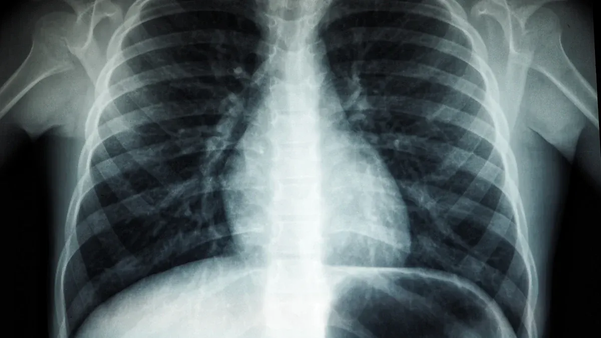
Identifying Abnormalities in the Chest or Lungs
X-rays are also commonly used to examine the chest and lungs. They can help identify abnormalities like tumors, fluid buildup, or other signs of lung cancer. This quick imaging method provides an initial look at potential issues that may require further investigation.
Advantages and Disadvantages
Advantages: Quick, Cost-Effective, Widely Available
X-rays offer several advantages. They are quick, often taking just a few minutes to complete. They are also cost-effective compared to other imaging methods, making them accessible to many patients. Additionally, X-ray machines are widely available in hospitals and clinics, ensuring you can get the imaging you need without long delays.
Disadvantages: Limited Detail for Soft Tissues, Radiation Exposure
Despite their benefits, X-rays have limitations. They provide limited detail when imaging soft tissues, which can make it harder to detect cancers in organs or muscles. Another concern is radiation exposure. Studies show that repeated exposure to X-rays can increase cancer risk, especially in children and adolescents. For this reason, doctors carefully weigh the benefits and risks before recommending X-rays.
Note: While X-rays play a crucial role in cancer diagnosis, they are often used alongside other imaging methods like CT scans or MRIs to provide a more comprehensive view.
How CT Scans Work and Their Applications in Cancer Diagnosis
How CT Scans Work
Combining X-rays with Computer Technology
CT scans, or computed tomography scans, use a combination of X-rays and computer technology to create detailed images of your body. Unlike traditional X-rays, CT scans take multiple images from different angles. A computer then combines these images to produce cross-sectional views of your organs and tissues. This process allows doctors to see structures inside your body with much greater clarity.
Role of Contrast Agents
Sometimes, doctors use contrast agents to improve the visibility of certain tissues or blood vessels during a CT scan. These agents, often injected into your bloodstream or swallowed, highlight specific areas of your body. For example, they can make tumors or abnormal growths stand out more clearly in the images.
Applications in Cancer Diagnosis
Detecting Tumors in Organs Like the Liver, Lungs, or Pancreas
CT scans are highly effective at detecting tumors in organs such as the liver, lungs, or pancreas. They provide detailed images that help doctors identify the size, shape, and location of a tumor. This makes CT scans a valuable tool for diagnosing cancers in these critical areas.
Assessing Cancer Spread and Staging
Doctors also use CT scans to determine if cancer has spread to other parts of your body. This process, known as staging, is essential for planning treatment. For example, CT scans can reveal whether lung cancer has metastasized to nearby lymph nodes or distant organs.
Did You Know? CT scans play a key role in tailoring treatment plans. They help doctors decide on the best approach based on the stage of cancer.
Advantages and Disadvantages
Advantages: Detailed Cross-Sectional Images, Useful for Staging
CT scans offer several advantages. They provide detailed cross-sectional images, allowing doctors to examine your body layer by layer. This level of detail is especially useful for staging cancer and monitoring its progression. Additionally, CT scans can quickly image large areas of your body, making them efficient for detecting metastases.
Disadvantages: Higher Radiation Exposure, Potential Contrast Agent Reactions
However, CT scans also have drawbacks. They expose you to higher levels of radiation compared to standard X-rays. Repeated exposure may increase your risk of developing other health issues. Contrast agents, while helpful, can sometimes cause allergic reactions or kidney problems in certain individuals. Doctors carefully evaluate these risks before recommending a CT scan.
Role in Cancer Detection | Contribution to Patient Care | |
|---|---|---|
X-rays | Non-invasive visualization of tumors | Early detection of treatable cancer |
CT scans | Accurate staging of cancer | Tailoring treatment plans based on cancer stage |
MRIs | Visualization of tumors in detail | Impacting treatment decisions and prognostic assessments |
Tip: CT scans are often preferred for diagnosing and staging cancers that affect large areas of the body, such as lung or abdominal cancers.
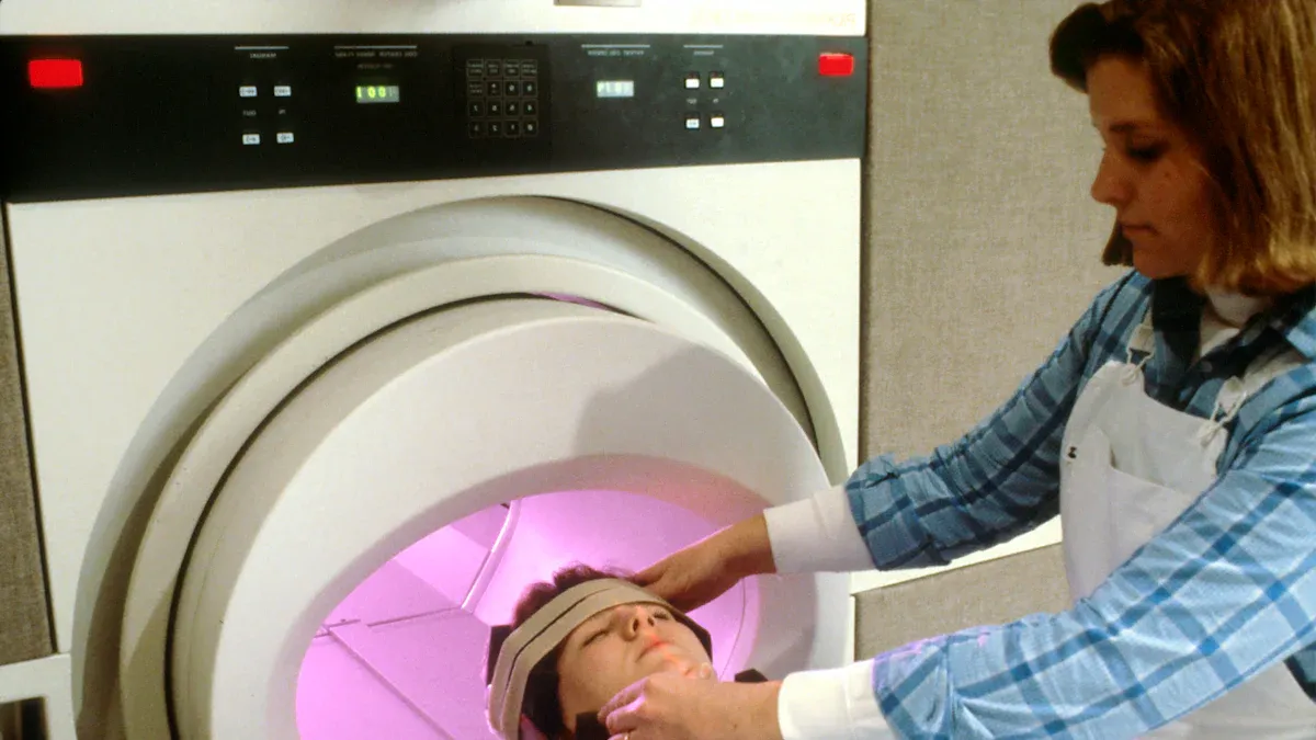
How MRIs Work and Their Applications in Cancer Diagnosis
How MRIs Work
Magnetic Fields and Radio Waves
MRIs, or magnetic resonance imaging, use powerful magnetic fields and radio waves to create detailed images of your body. The magnetic field aligns hydrogen atoms in your body, while radio waves disrupt this alignment. When the atoms return to their original position, they release energy. The MRI machine captures this energy and converts it into images. This process allows doctors to see inside your body without using radiation.
Superior Imaging for Soft Tissues
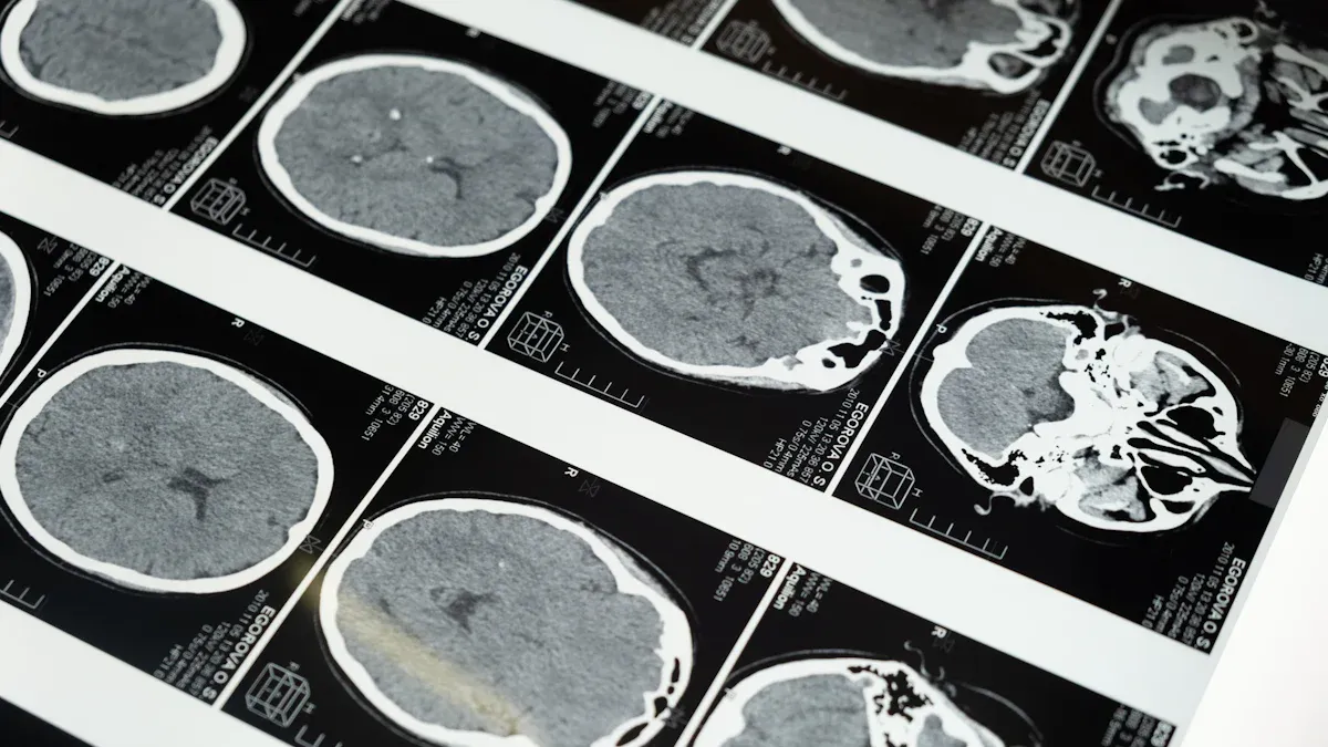
MRIs excel at imaging soft tissues. They provide clear and detailed views of organs, muscles, and other soft structures. This makes them especially useful for detecting abnormalities that other imaging methods might miss. For example, MRIs can reveal subtle changes in the brain or spinal cord that are critical for diagnosis.
Tip: If you feel nervous about the confined space of an MRI machine, ask your doctor about open MRI options or relaxation techniques.
Applications in Cancer Diagnosis
Detecting Brain, Spinal Cord, and Soft Tissue Cancers
Doctors often use MRIs to detect cancers in the brain, spinal cord, and soft tissues. These scans can identify tumors, pinpoint their location, and determine their size. For example, an MRI can help diagnose brain tumors or sarcomas in muscles. This level of detail is crucial for planning treatment.
Monitoring Treatment Progress
MRIs also play a key role in monitoring how well your treatment is working. They can track changes in tumor size or detect new growths. This helps doctors adjust your treatment plan as needed, ensuring the best possible outcome.
Advantages and Disadvantages
Advantages: No Radiation, Excellent Soft Tissue Detail
One major advantage of MRIs is that they do not use radiation. This makes them safer for repeated use, especially for children or individuals requiring frequent scans. Additionally, MRIs provide unmatched detail for soft tissues, making them ideal for diagnosing complex conditions.
Disadvantages: Expensive, Time-Consuming, Not Suitable for Patients with Metal Implants
Despite their benefits, MRIs have some drawbacks. They are more expensive than other imaging methods, which can limit accessibility. The scans also take longer, often lasting 30 minutes to an hour. Furthermore, MRIs are not suitable for patients with metal implants, such as pacemakers, due to the strong magnetic field.
Note: Always inform your doctor about any implants or medical devices before undergoing an MRI.
Comparing X-rays, CT Scans, and MRIs
Key Differences
Imaging Technology and Mechanisms
Each imaging method uses unique technology to capture internal images of your body. X-rays rely on electromagnetic radiation to create shadow-like images, which are ideal for visualizing dense tissues like bones. CT scans take this a step further by combining X-rays with computer technology to produce detailed cross-sectional images. MRIs, on the other hand, use magnetic fields and radio waves to generate highly detailed images, especially of soft tissues. These differences make each method suitable for specific diagnostic purposes.
Types of Tissues and Cancers They Are Best Suited For
X-rays work best for detecting abnormalities in bones or the chest, such as bone cancers or lung tumors. CT scans excel at imaging organs like the liver, pancreas, or lungs, making them valuable for diagnosing and staging cancers in these areas. MRIs are unmatched when it comes to soft tissues, making them the preferred choice for detecting brain tumors, spinal cord cancers, or sarcomas.
Similarities
Non-Invasive Diagnostic Tools
All three imaging methods are non-invasive, meaning they do not require surgery or incisions. This makes them safer and more comfortable for you during the diagnostic process.
Essential for Cancer Detection and Monitoring
These tools play a critical role in cancer care. They help doctors detect cancer early, determine its stage, and monitor how well treatments are working. For example, "The Role of Imaging in Cancer Diagnosis: X-Rays" highlights how X-rays can provide quick insights into bone metastases or lung abnormalities.
Choosing the Right Imaging Method
Factors Doctors Consider, Such as Cancer Type and Patient Condition
Doctors choose the imaging method based on your specific needs. They consider the type of cancer, its location, and your overall health. For instance, if you have a suspected brain tumor, an MRI might be the best option due to its superior soft tissue detail.
Balancing Accuracy, Safety, and Cost
Doctors also weigh the benefits and risks of each method. X-rays and CT scans involve radiation exposure, while MRIs are radiation-free but more expensive. They aim to balance accuracy, safety, and cost to ensure you receive the most effective care.
Tip: Ask your doctor to explain why a specific imaging method is recommended for your condition. This can help you feel more informed and confident about your care.
X-rays, CT scans, and MRIs each serve unique purposes in cancer diagnosis. X-rays provide quick insights into dense tissues like bones, while CT scans offer detailed cross-sectional images for staging cancers. MRIs excel at capturing soft tissue details without radiation. Doctors choose the best imaging method based on your cancer type, body area, and diagnostic needs. Understanding these differences helps you engage in informed discussions with your healthcare provider, empowering you to make confident decisions about your care.
Tip: Ask your doctor how each imaging method contributes to your diagnosis and treatment plan.
FAQ
What should you expect during an X-ray, CT scan, or MRI procedure?
X-ray: You’ll stand or lie still while the machine takes images.
CT scan: You’ll lie on a table that moves through a scanner.
MRI: You’ll lie inside a tube-like machine, which may feel confined.
Tip: Wear comfortable clothing and remove metal objects before your scan.
Are these imaging methods safe for children?
Yes, doctors use these methods cautiously for children. They minimize radiation exposure by adjusting the settings for X-rays and CT scans. MRIs, which don’t use radiation, are often preferred for younger patients.
Note: Always discuss safety concerns with your child’s doctor before the procedure.
How long does each imaging test take?
X-ray: A few minutes.
CT scan: 10-30 minutes.
MRI: 30-60 minutes or longer, depending on the area being scanned.
Tip: Bring a book or music for longer procedures like MRIs to stay relaxed.
Can you eat or drink before these scans?
It depends on the scan. For X-rays, no preparation is needed. CT scans and MRIs may require fasting if contrast agents are used. Your doctor will provide specific instructions.
Reminder: Follow all pre-scan guidelines to ensure accurate results.
What if you feel anxious about the scan?
Feeling anxious is normal, especially for MRIs. You can ask your doctor about open MRI options or mild sedatives. Relaxation techniques, like deep breathing, can also help.
Tip: Inform the technician about your concerns. They can guide you through the process.
See Also
Recognizing Duodenal Cancer: Key Symptoms And Treatment Options
Conjunctival Melanoma: Identifying Symptoms And Available Treatments
An In-Depth Overview Of Various Cancer Types Explained
Astrocytoma Overview: Exploring Its Various Types And Features

© 2026 Banish Cancer. Banish Cancer is a registered non-profit organization providing evidence-based research, educational courses, and Fear Response AI support services. Reg. No: 305706884 | Address: Taikos pr. 43-603, Kaunas, Lithuania.